Histology (microscopic anatomy)
Tissue science on real slides: high-resolution light micrographs with toggleable labels, identification tips and self-tests – arranged as a learning path from epithelium to nervous tissue.
1 Epithelial tissue
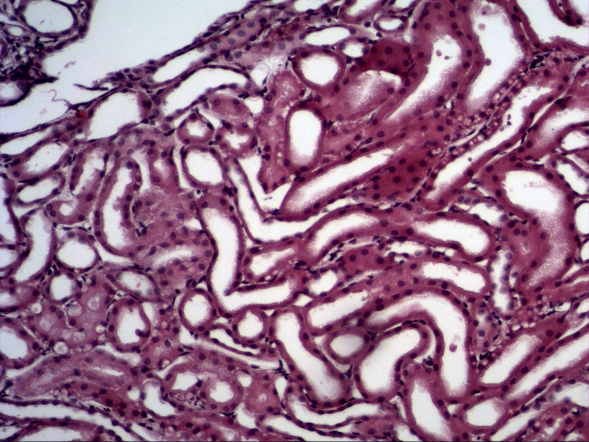
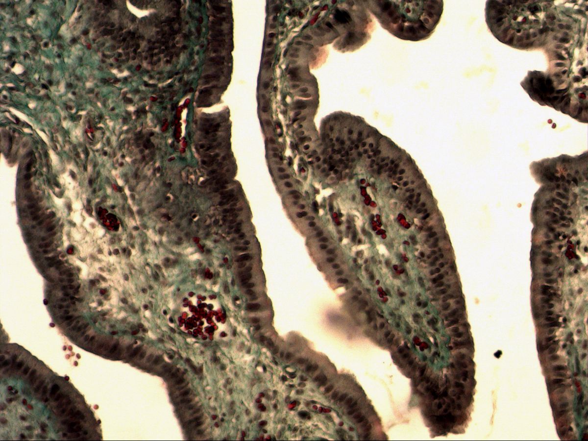
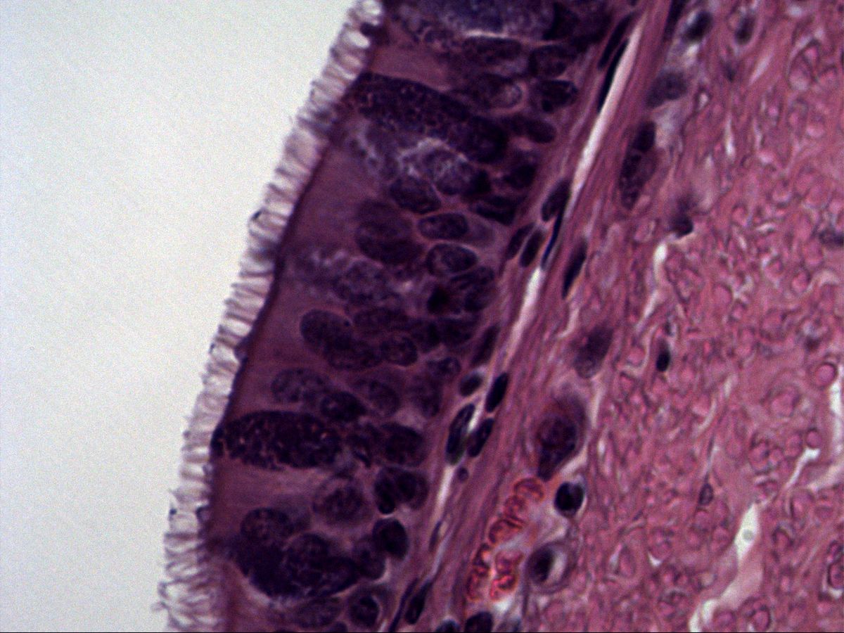
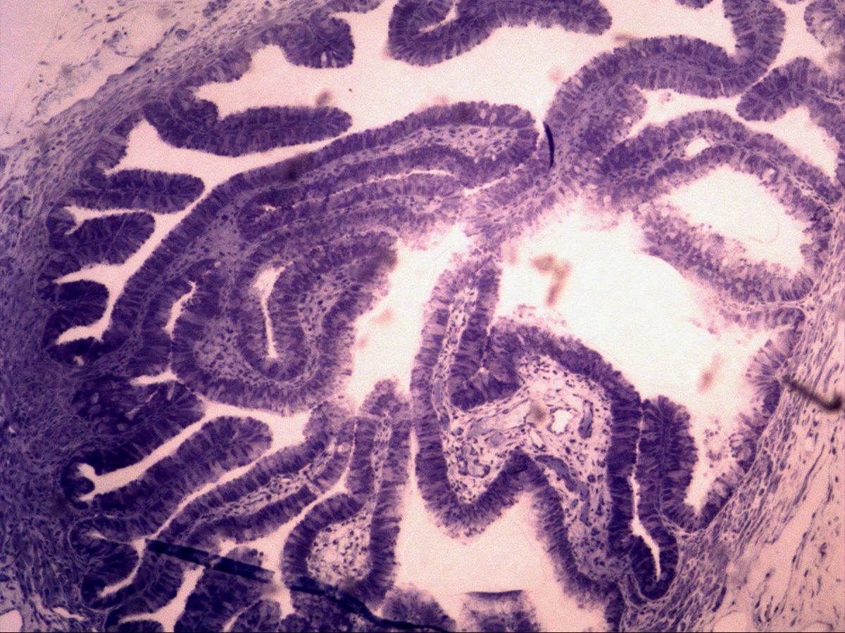
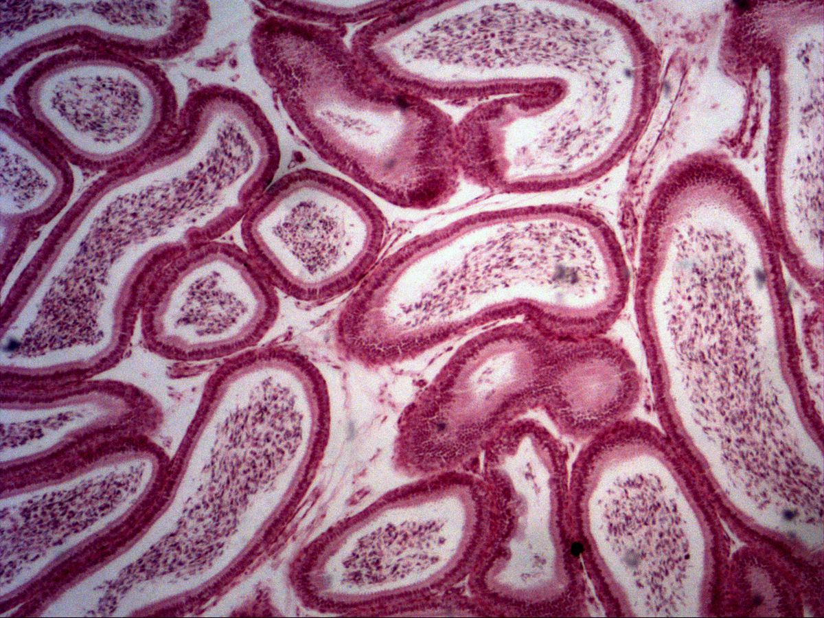
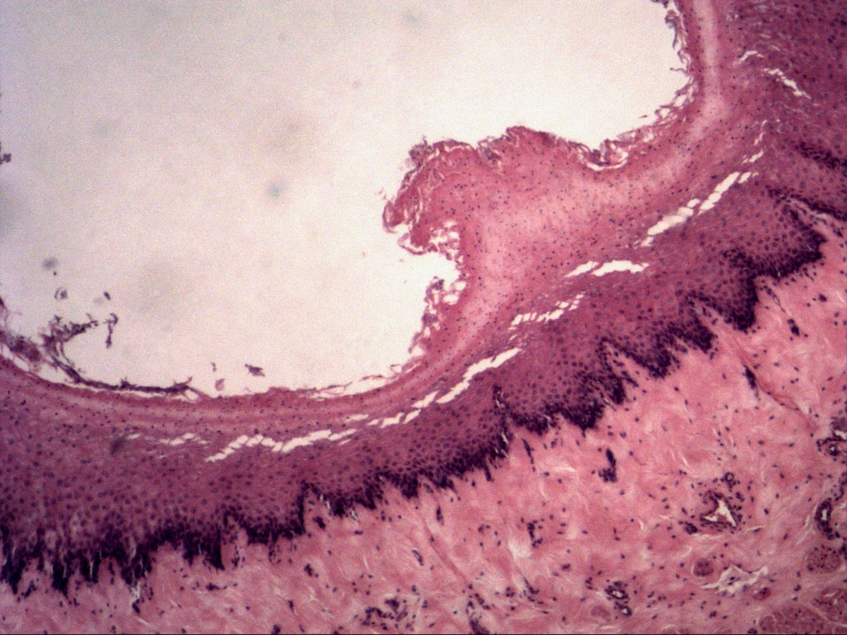
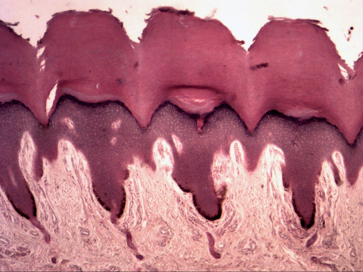
8 topicsEpithelia cover outer and inner surfaces. Learn to classify them reliably by layering (simple, pseudostratified, stratified), cell shape (squamous, cuboidal, columnar) and apical specialisations such as microvilli, cilia and stereocilia.
2 Glandular tissue
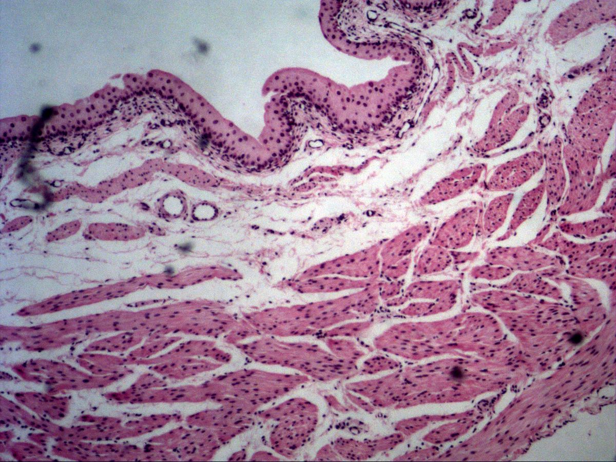
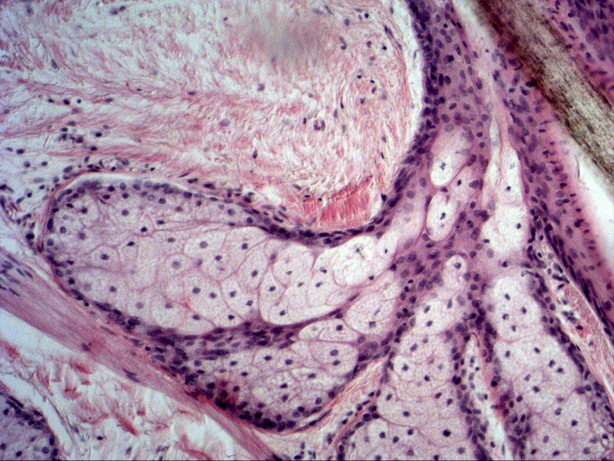
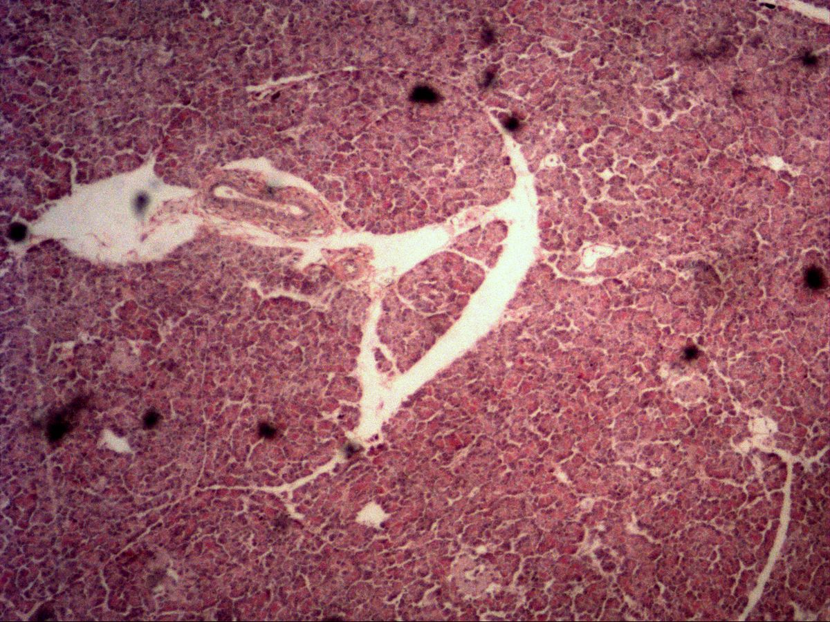
5 topicsFrom the single goblet cell to the islet of Langerhans: recognise exocrine and endocrine glands, their secretory end pieces, ducts and modes of secretion (merocrine, apocrine, holocrine) on real slides.
3 Germ cells & embryonic tissue
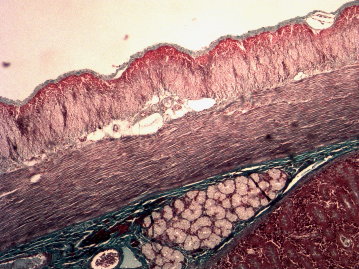
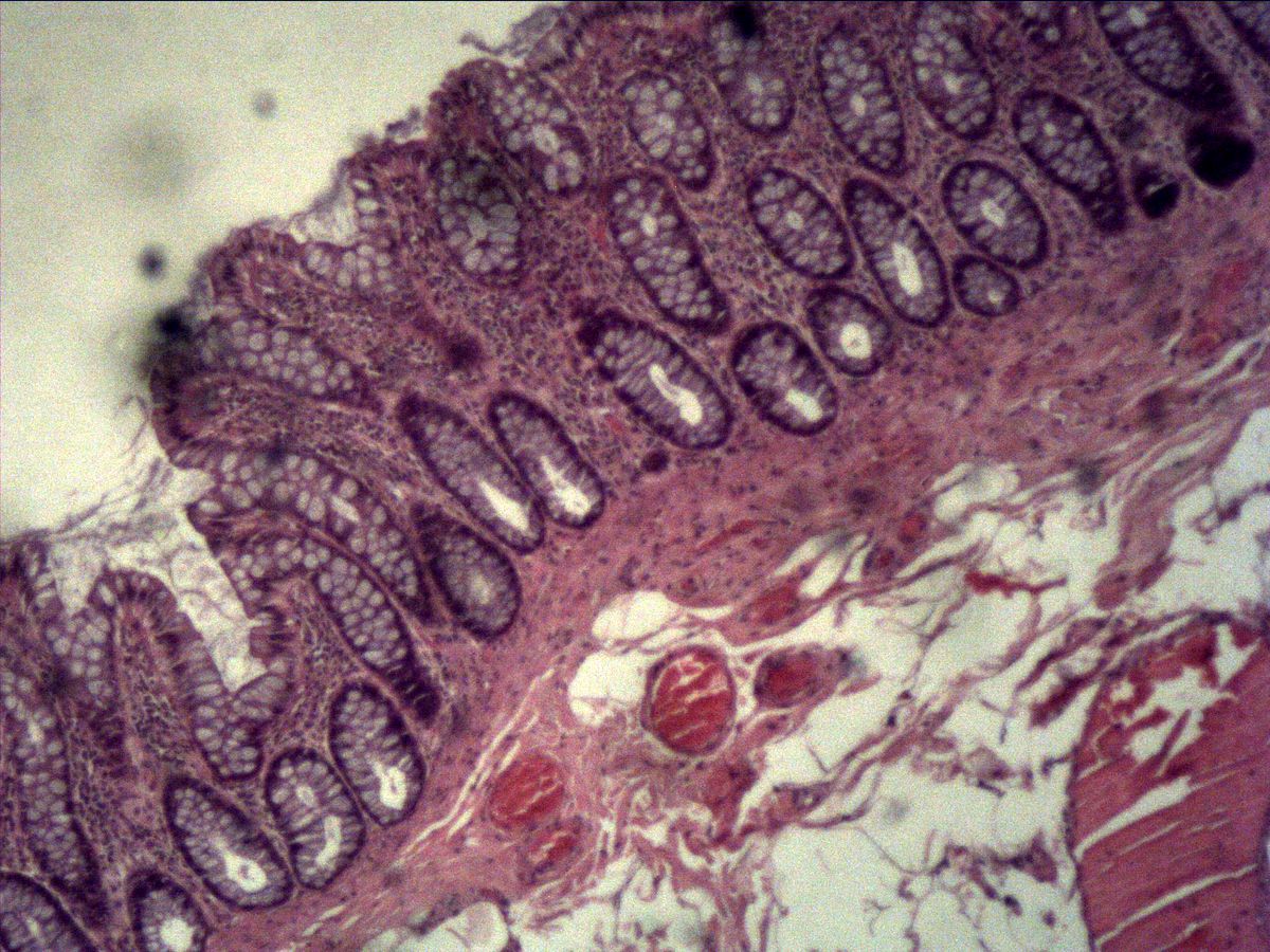
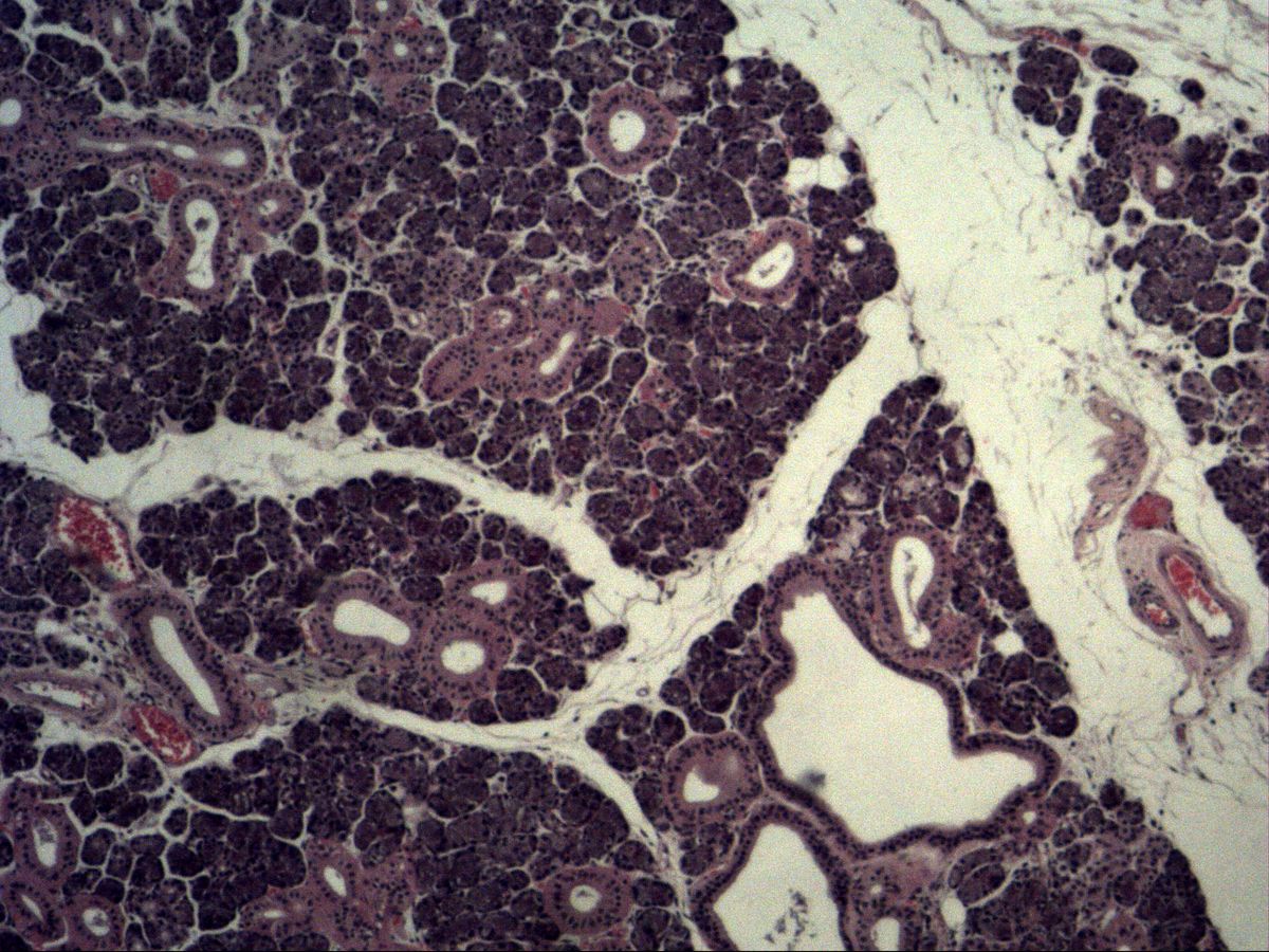
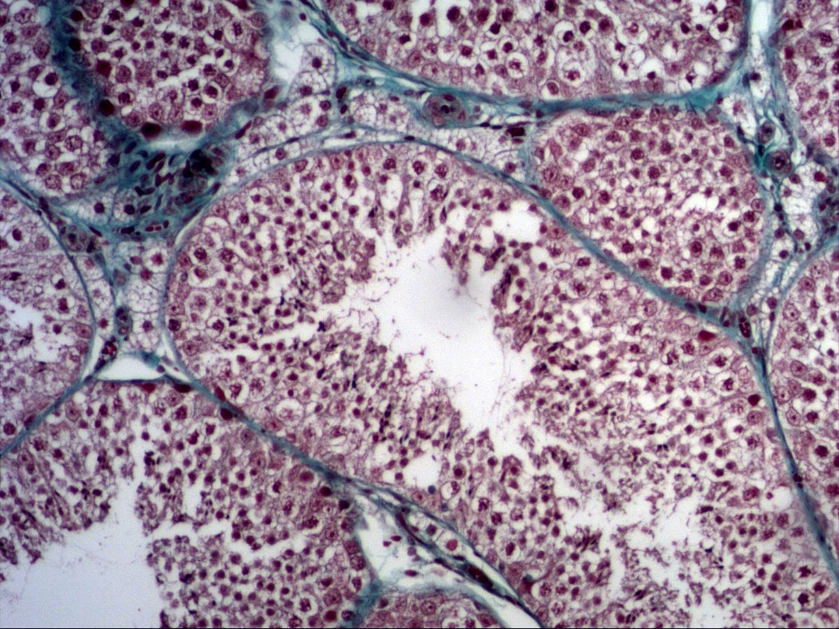
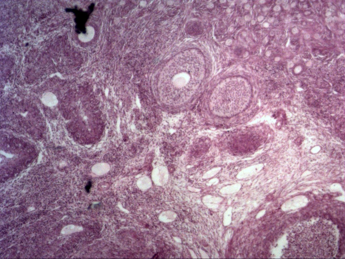
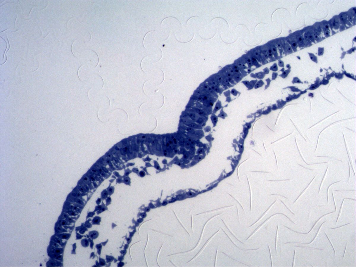
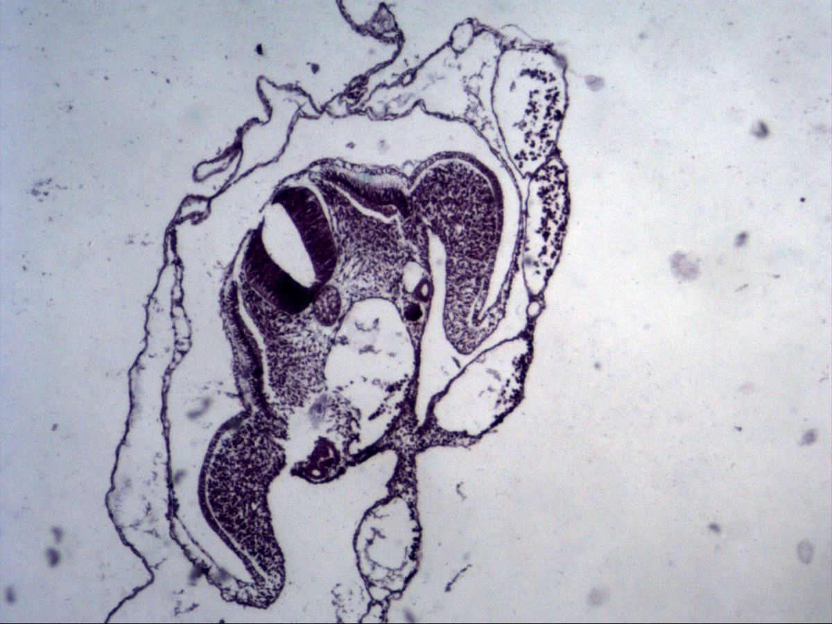
4 topicsSpermatogenesis in the seminiferous tubule, follicle maturation in the ovary and early embryonic development with gastrulation and neurulation – the basics of the germ layers in microscopic images.
4 Connective tissue
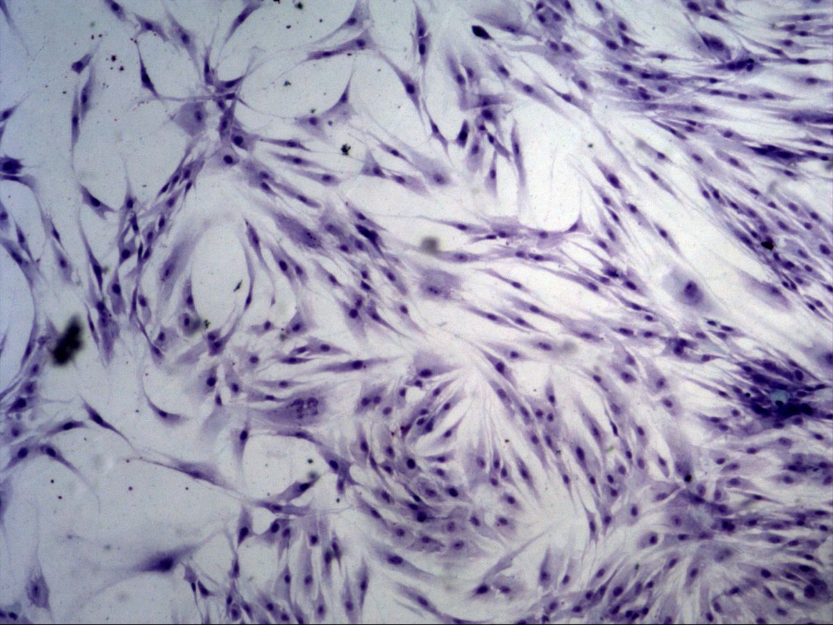
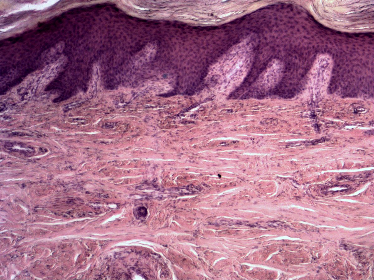
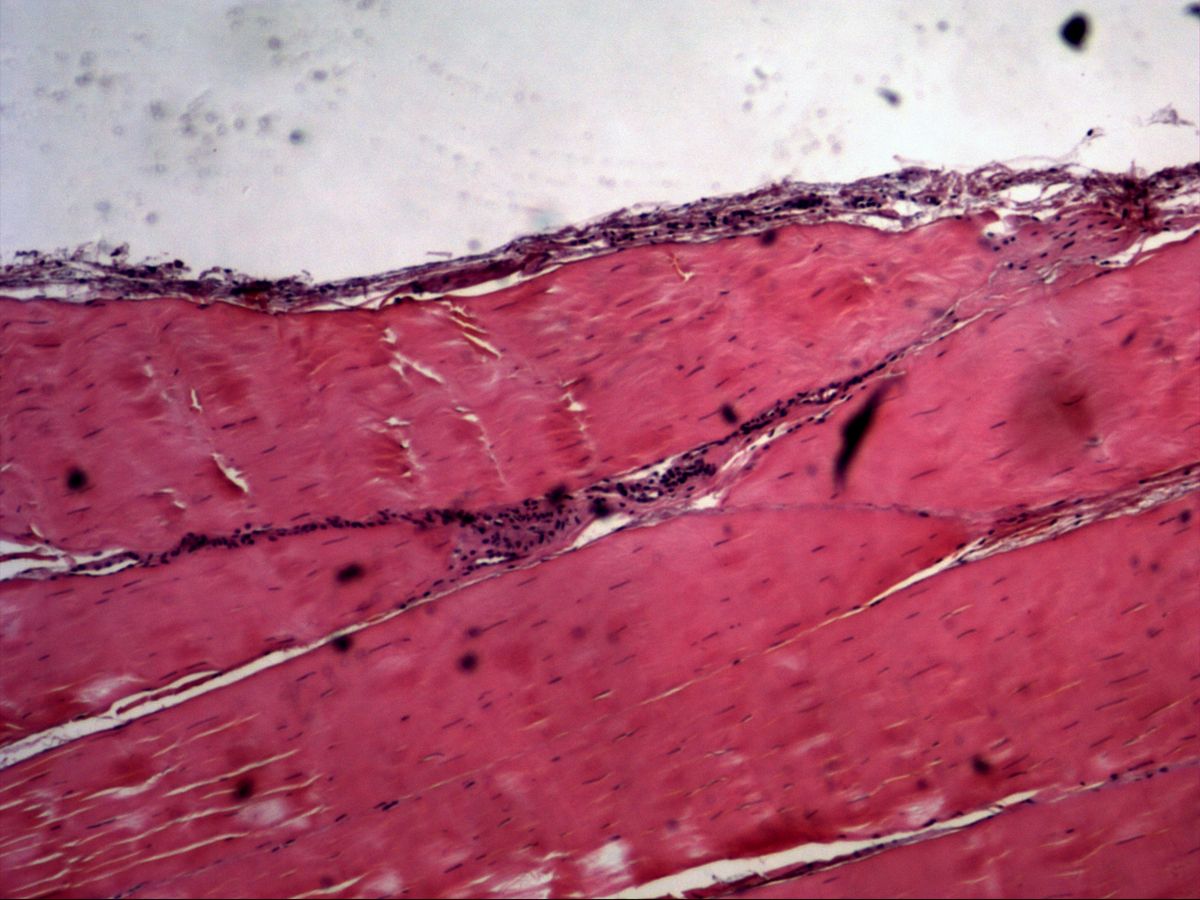
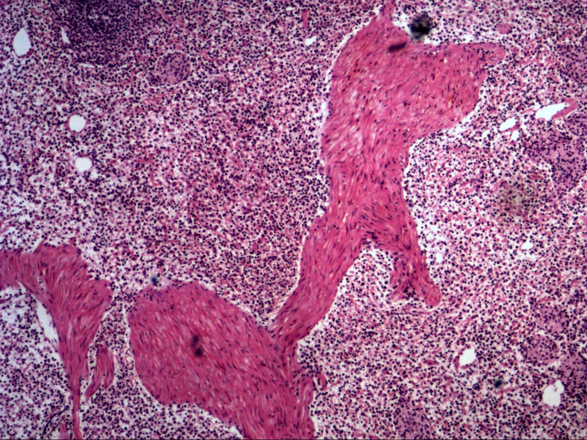
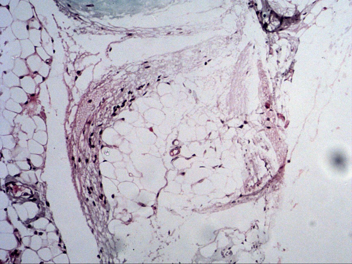
6 topicsCells and fibres working together: mucous, loose, dense, reticular connective tissue and adipose tissue – with fibroblasts, collagen and reticular fibres and adipocytes.
5 Cartilage & bone
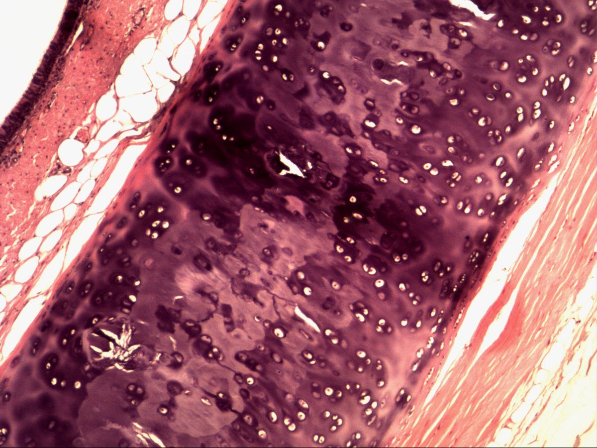
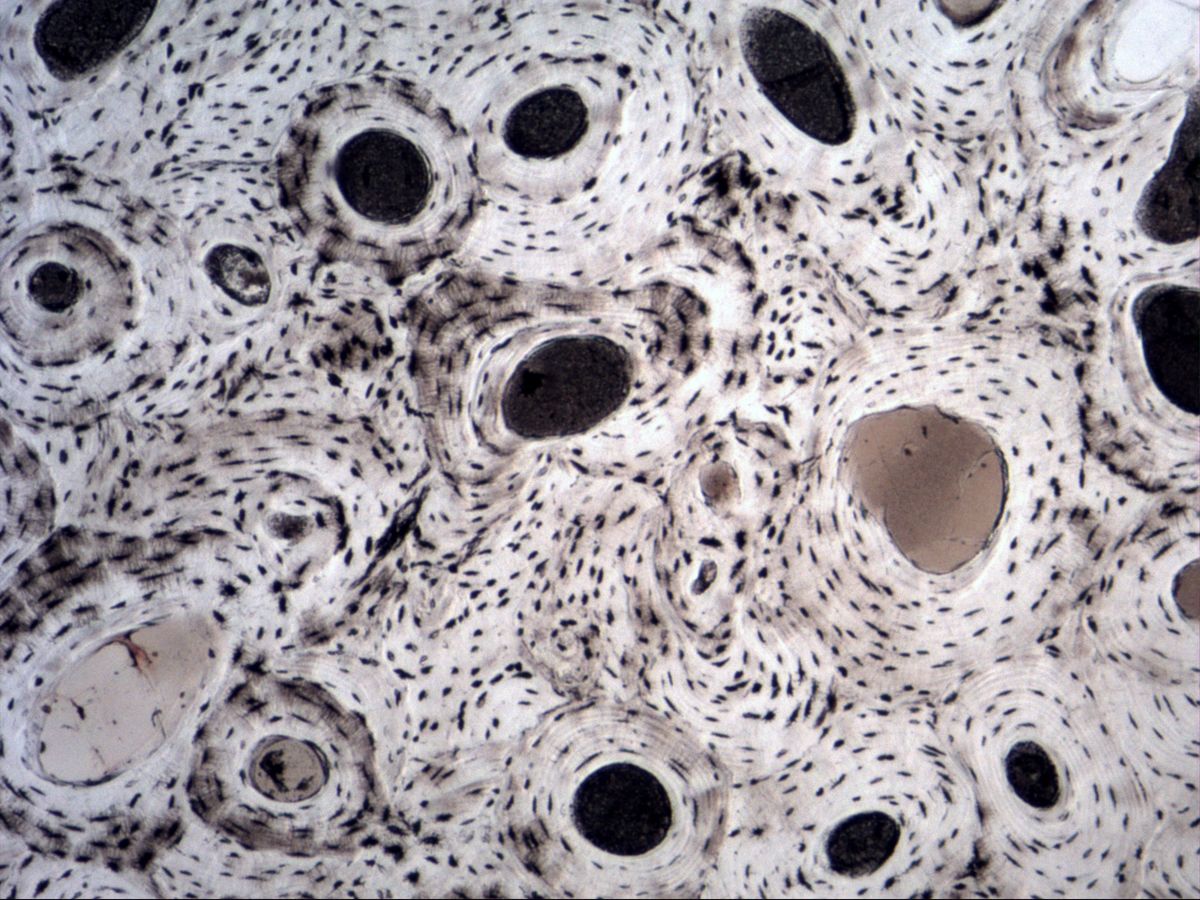
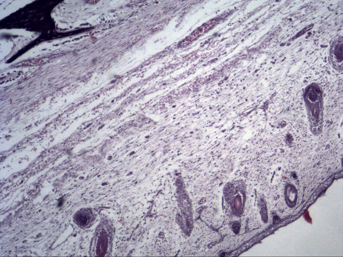
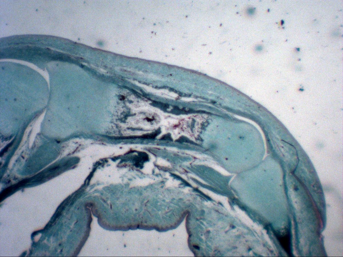
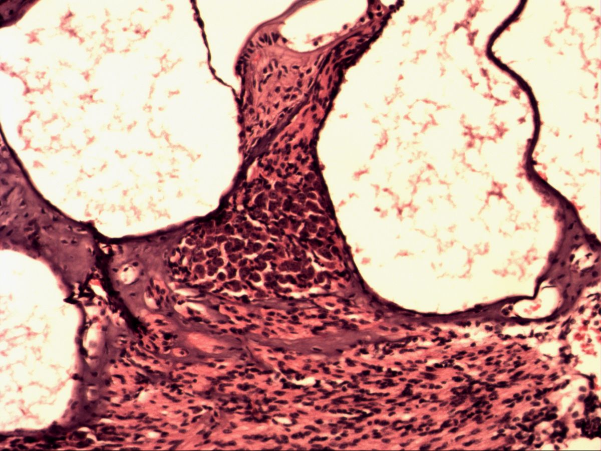
4 topicsHyaline cartilage, lamellar bone with osteons and the two routes of bone formation – intramembranous and endochondral ossification – step by step on real slides.
6 Muscle tissue
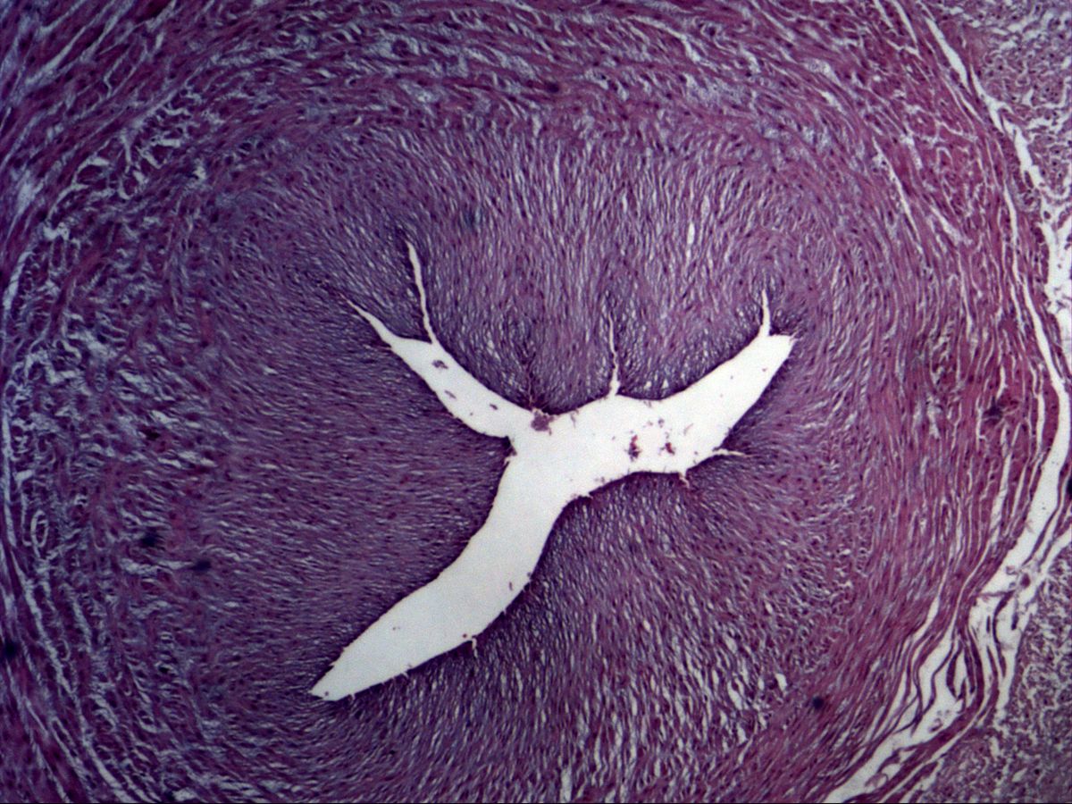
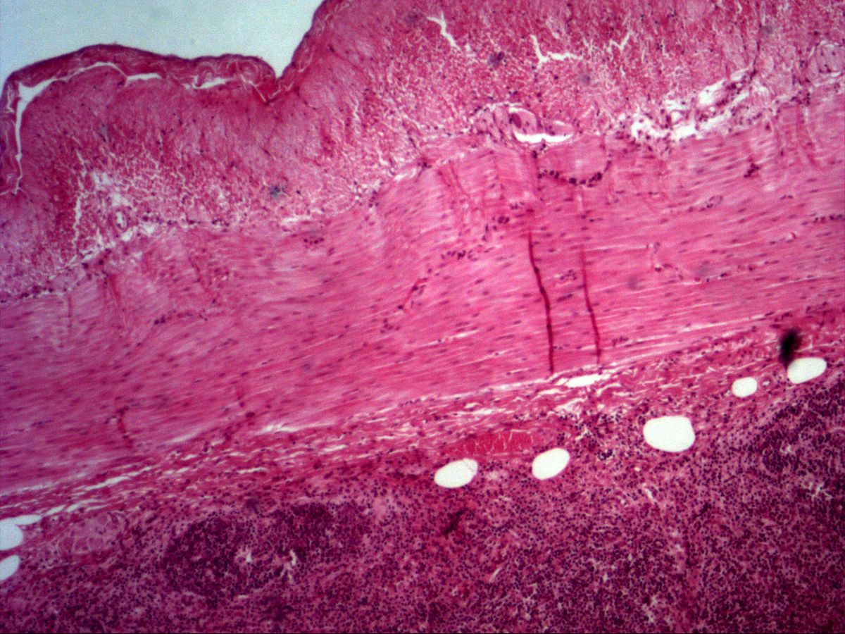
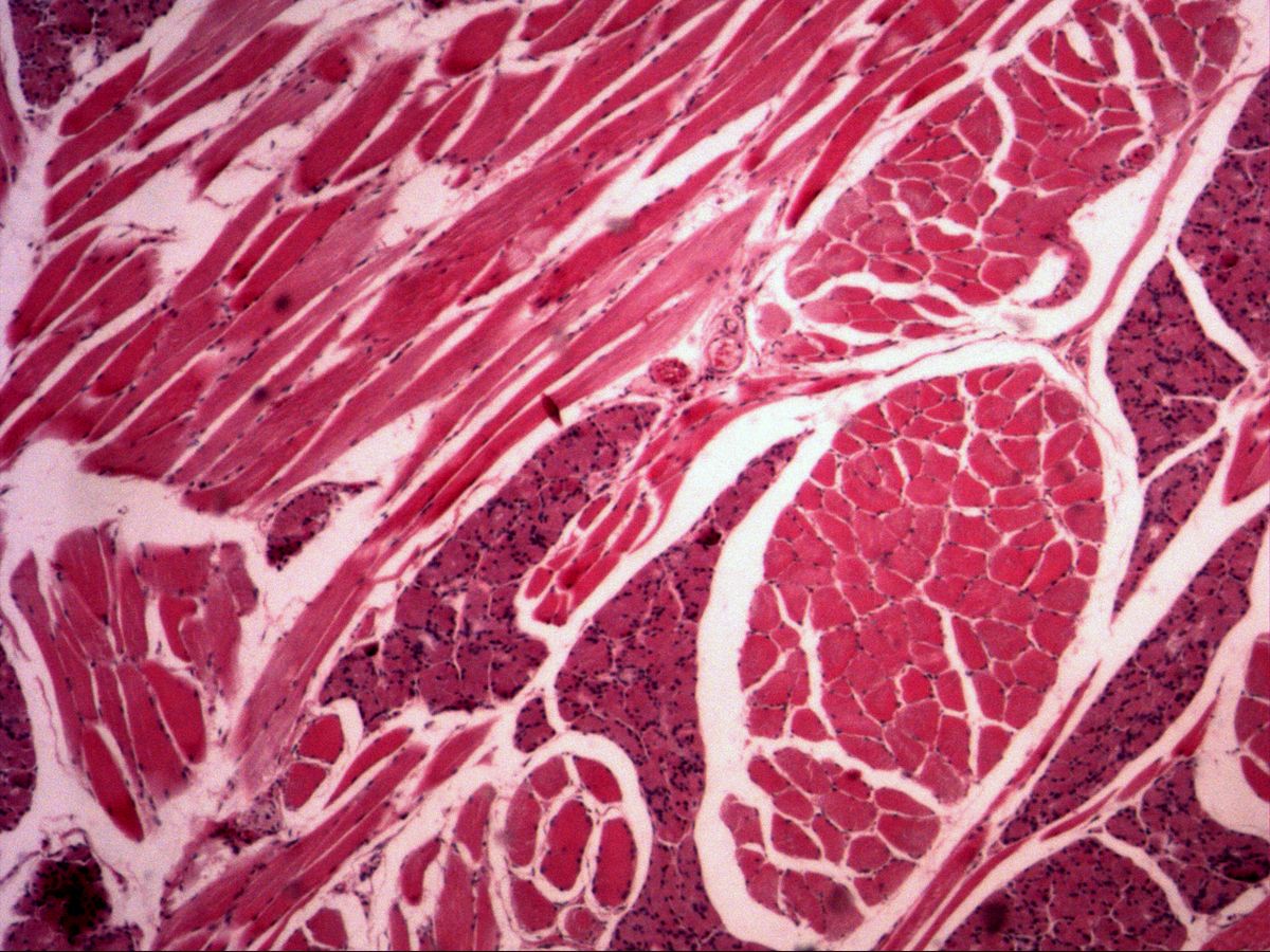
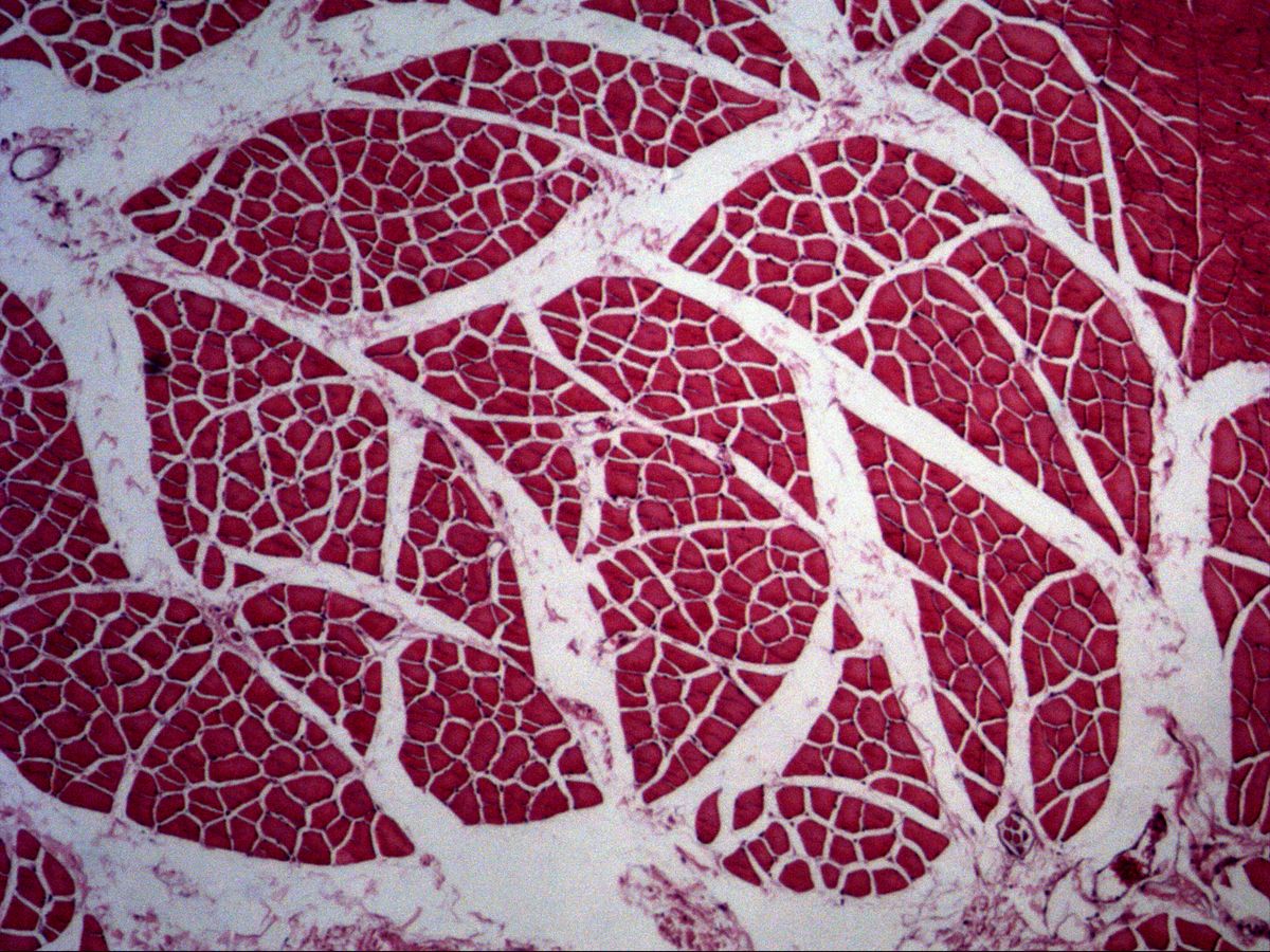
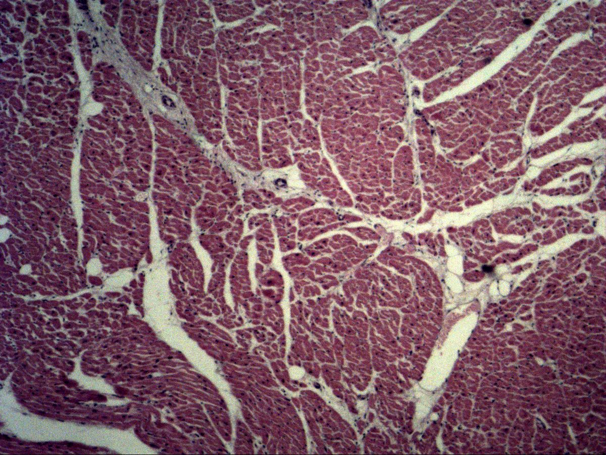
4 topicsDistinguish smooth, skeletal and cardiac muscle in longitudinal and cross-section: nucleus position, cross-striation, intercalated discs and connective-tissue sheaths.
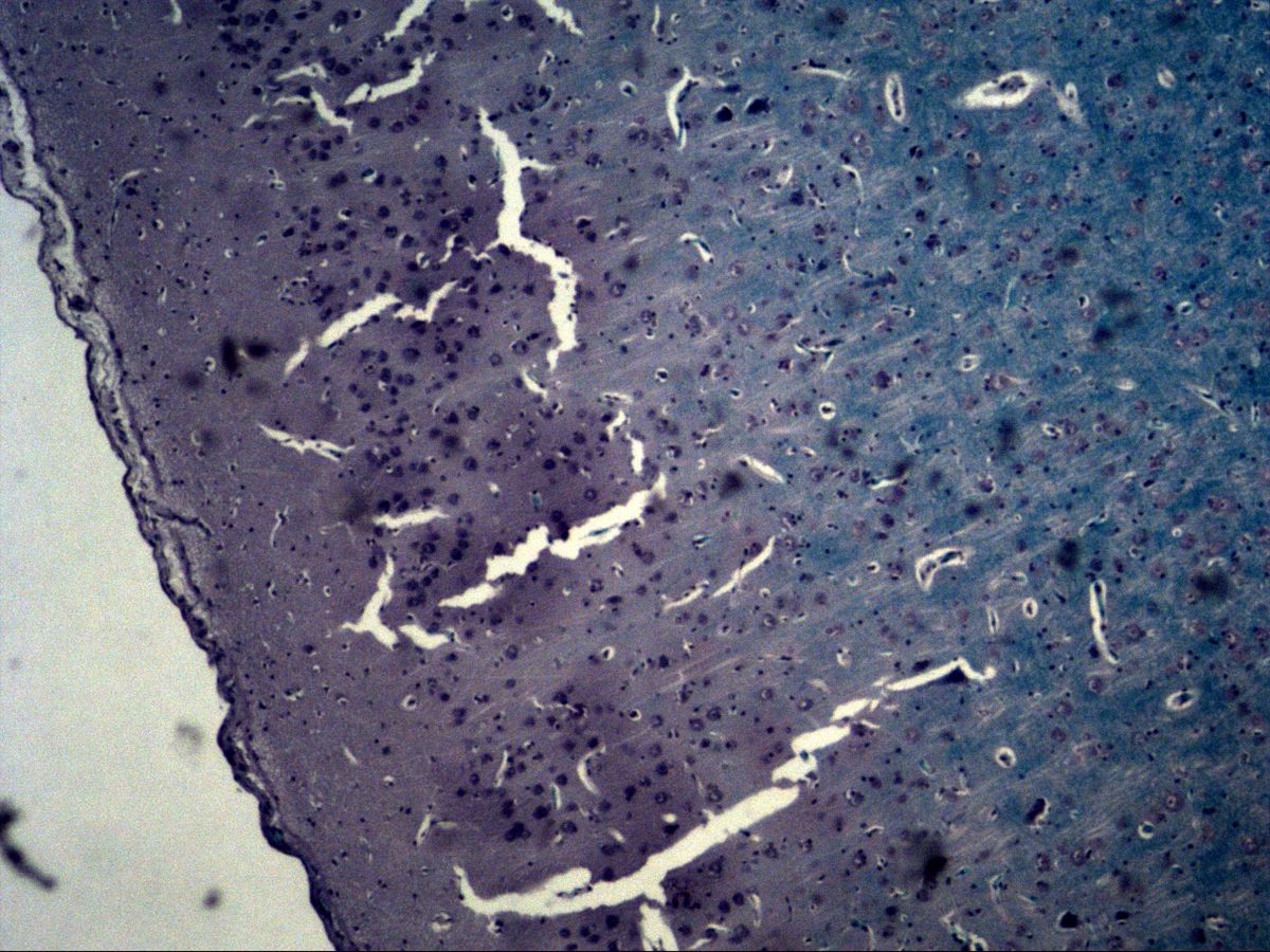
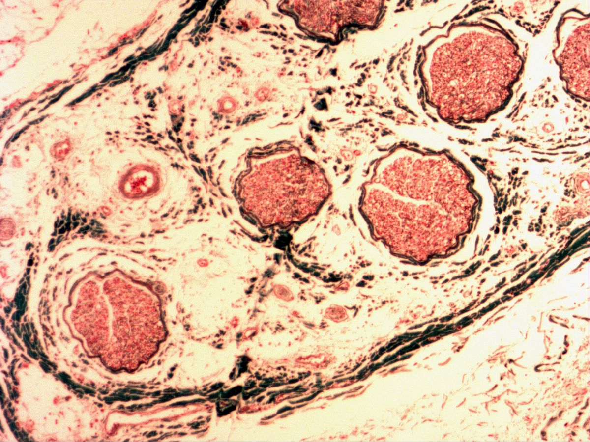
7 Nervous tissue
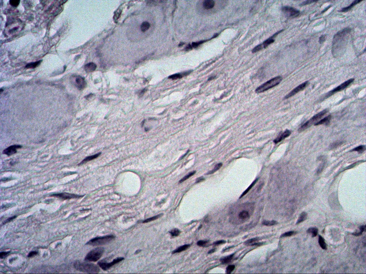
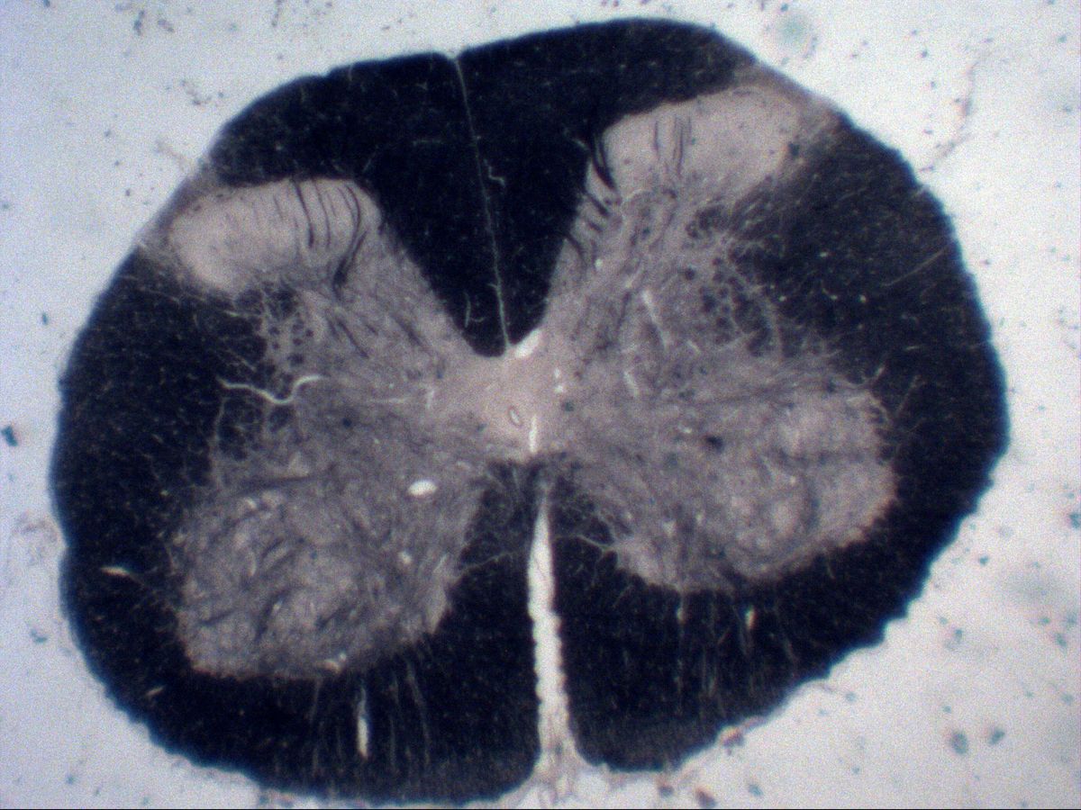
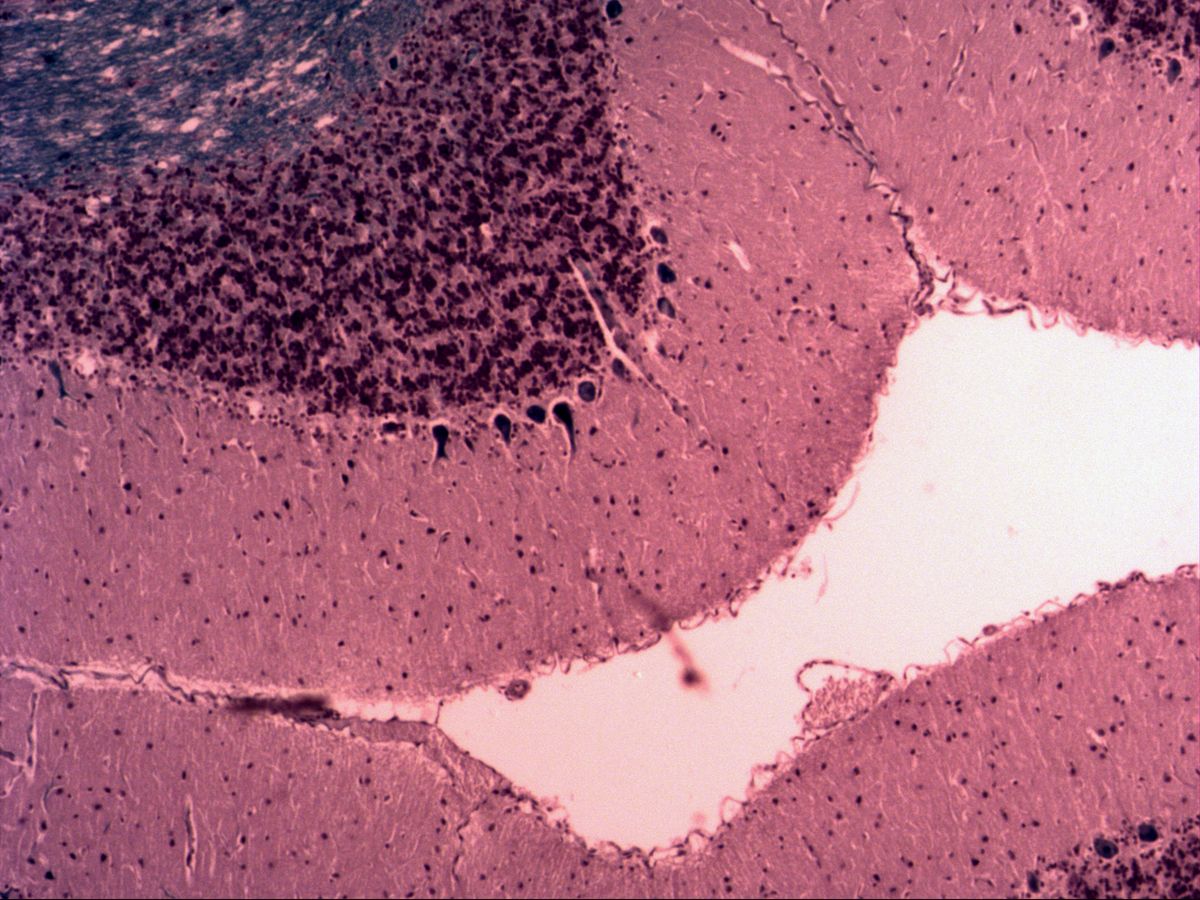
6 topicsPseudounipolar, bipolar and multipolar neurons, Purkinje and pyramidal cells and the structure of peripheral nerves – with glia, Nissl substance and myelin sheaths.
8 Lymphoid tissue
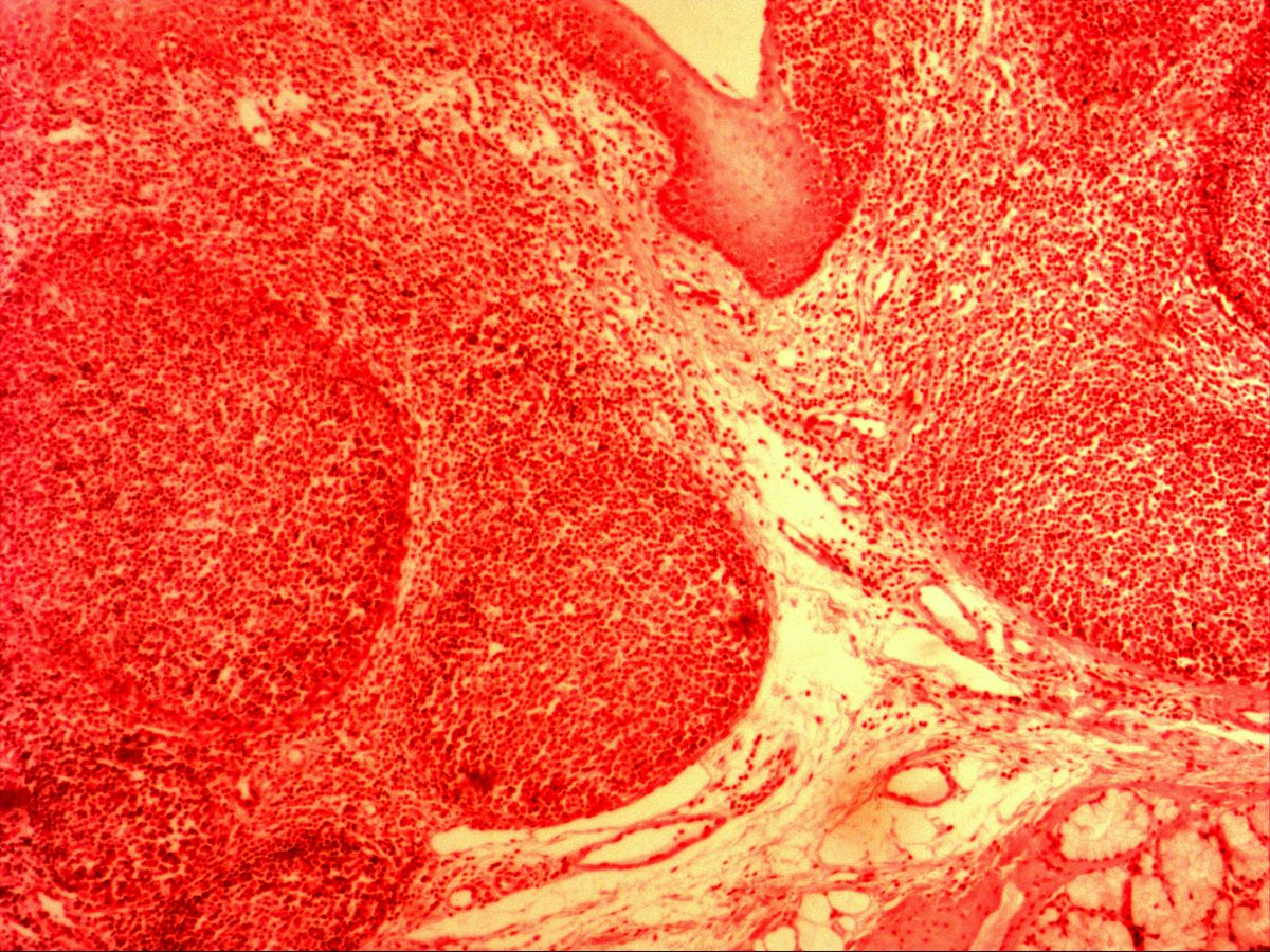
1 topicLymphoid follicles with germinal centre and mantle zone, lymphocytes and the framework of lymphoid organs.
9 Sensory organs
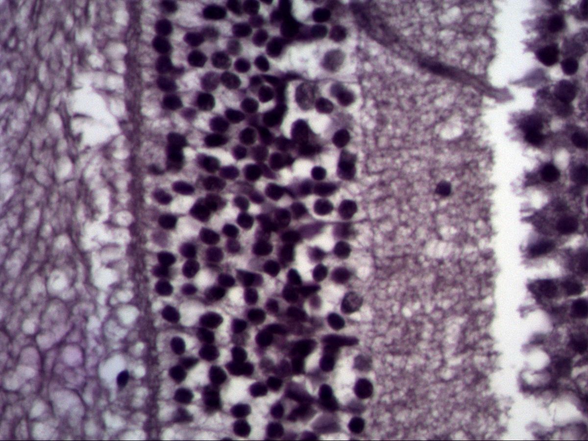
1 topicMicroscopic anatomy of the sense organs – starting with the layered structure of the retina.