Cardiac muscle
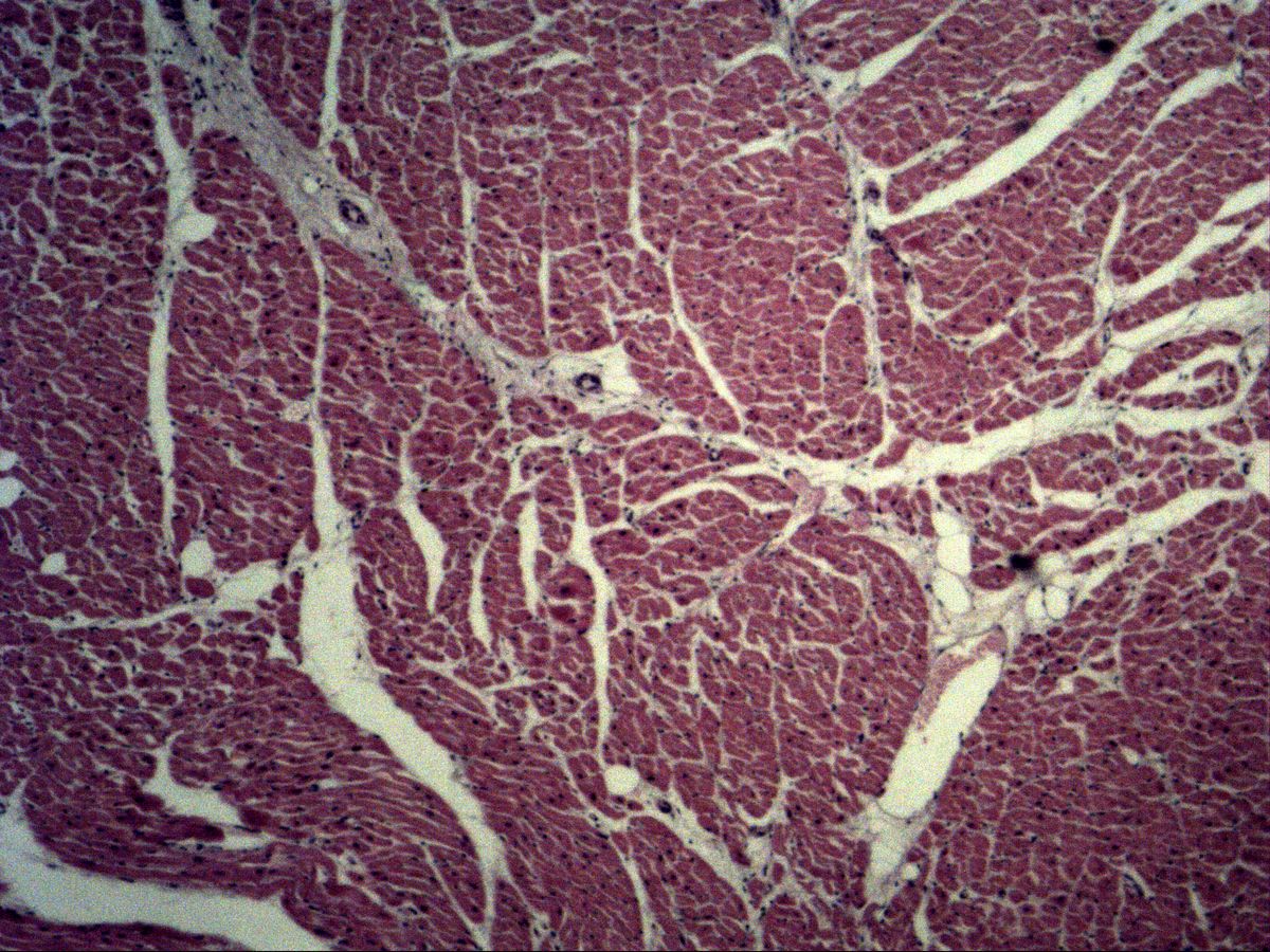
Myocardium · H&E
Labelled structures
This image has no labels – use the caption instead.
The myocardium consists of cardiac muscle cells (cardiomyocytes), which are striated like skeletal muscle but have a centrally placed nucleus, branch and are joined into a functional network by intercalated discs. In sections the cell bundles run in different directions, so longitudinally, obliquely and transversely cut areas lie side by side.
How to recognise it
- Cross-striation as in skeletal muscle
- One (rarely two) central nucleus per cell
- Perinuclear, myofibril-free halo
- Branched cells
- Intercalated discs
- Highly capillarised interstitium
Structures to find
- Centrally placed nucleus
- Perinuclear fibril-free zone
- Branching of a cardiomyocyte
- Intercalated disc
- Cross-striation
- Capillaries with erythrocytes
Structure
Cardiomyocytes are about 100 µm long and 10–20 µm thick and usually have one, occasionally two central oval nuclei. At the nuclear poles is a myofibril-free, pale zone (perinuclear halo) containing mitochondria, Golgi apparatus and lipofuscin. The cells branch in a Y-shape and are joined end to end by intercalated discs – step-like lines across the fibre containing adherens junctions, desmosomes and gap junctions. Between the cells lies loose connective tissue with a very dense capillary network.
Function
Cardiac muscle contracts rhythmically, involuntarily and without fatigue. Excitation arises in the sinoatrial node and spreads from cell to cell via the gap junctions of the intercalated discs – the myocardium works as a functional syncytium. The high energy demand is met by abundant mitochondria and dense capillary supply. Adult cardiomyocytes hardly divide; lost tissue is replaced by scar tissue.
Microscopy tips
Look for a longitudinally cut area. Watch for one nucleus per cell in the middle of the fibre, often with a pale halo at both poles, and for branching fibres. In H&E, intercalated discs can only be guessed at high magnification as fine darker transverse lines; with iron haematoxylin they stand out clearly. In cross-section, the nuclei lie centrally in roundish, irregular profiles. Differential diagnosis: skeletal muscle has peripheral nuclei and unbranched fibres; smooth muscle lacks striation and has spindle-shaped cells.
Clinical relevance
In myocardial infarction, cardiomyocytes die and are replaced within weeks by collagen-rich scar tissue. Under chronic pressure overload (e.g. hypertension), the myocardium responds with hypertrophy: the cells thicken and their nuclei become large and plump. In hypertrophy the number of capillaries does not increase to the same extent, so the myocardium becomes more vulnerable to oxygen shortage. Histologically, an infarct is dated by cell necrosis, neutrophil infiltration, granulation tissue and finally scar.
Slide images
Test your knowledge
Assign the correct number in the image to each term.
Tap the requested structure in the image.
Multiple-choice questions on structure and function.
Frequently asked questions
What are intercalated discs?
Step-like cell junctions between cardiomyocytes. They contain adherens junctions and desmosomes for mechanical coupling and gap junctions for electrical conduction.
Why are intercalated discs hard to see in H&E?
They are very thin and stain little differently from the sarcoplasm in H&E. At high magnification and careful focusing they appear as fine dark transverse lines; iron haematoxylin shows them better.
How do I distinguish cardiac from skeletal muscle?
Cardiomyocytes have a central nucleus, branch and have intercalated discs. Skeletal muscle fibres are unbranched with many peripheral nuclei.
Related topics
Updated: