Learning path · Histology
Cartilage & bone
Hyaline cartilage, lamellar bone with osteons and the two routes of bone formation – intramembranous and endochondral ossification – step by step on real slides.
-
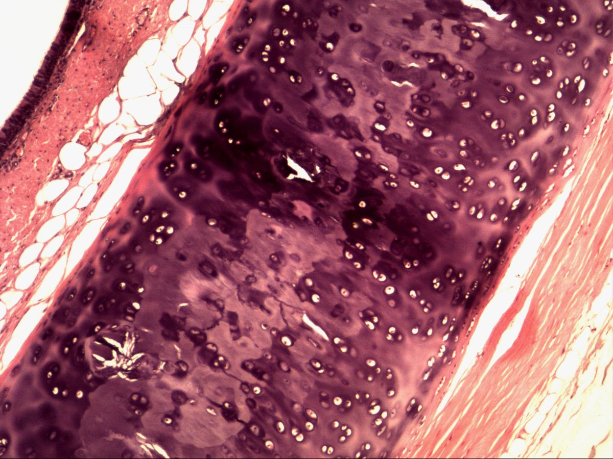
Hyaline cartilage
Trachea · H&E
5 images 14 structures -
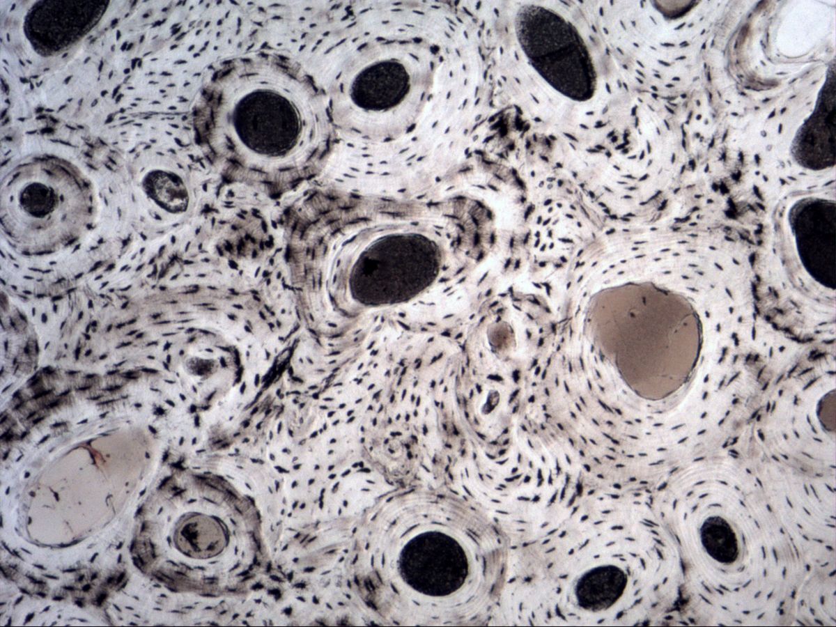
Lamellar bone
Tibia · ground section
6 images 13 structures -
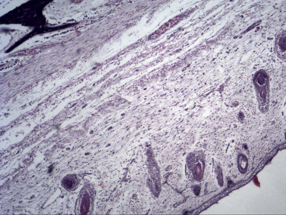
Intramembranous ossification
Fetal tissue · H&E
3 images 14 structures -
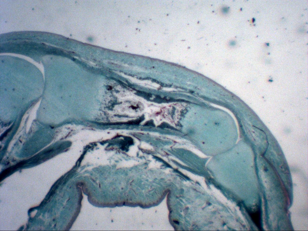
Endochondral ossification
Fetal phalanx · trichrome
9 images 19 structures
Test your knowledge
Mixed exam simulation: identify slides, name highlighted structures and answer knowledge questions. Choose your areas and off you go.