Hyaline cartilage
Trachea · H&E
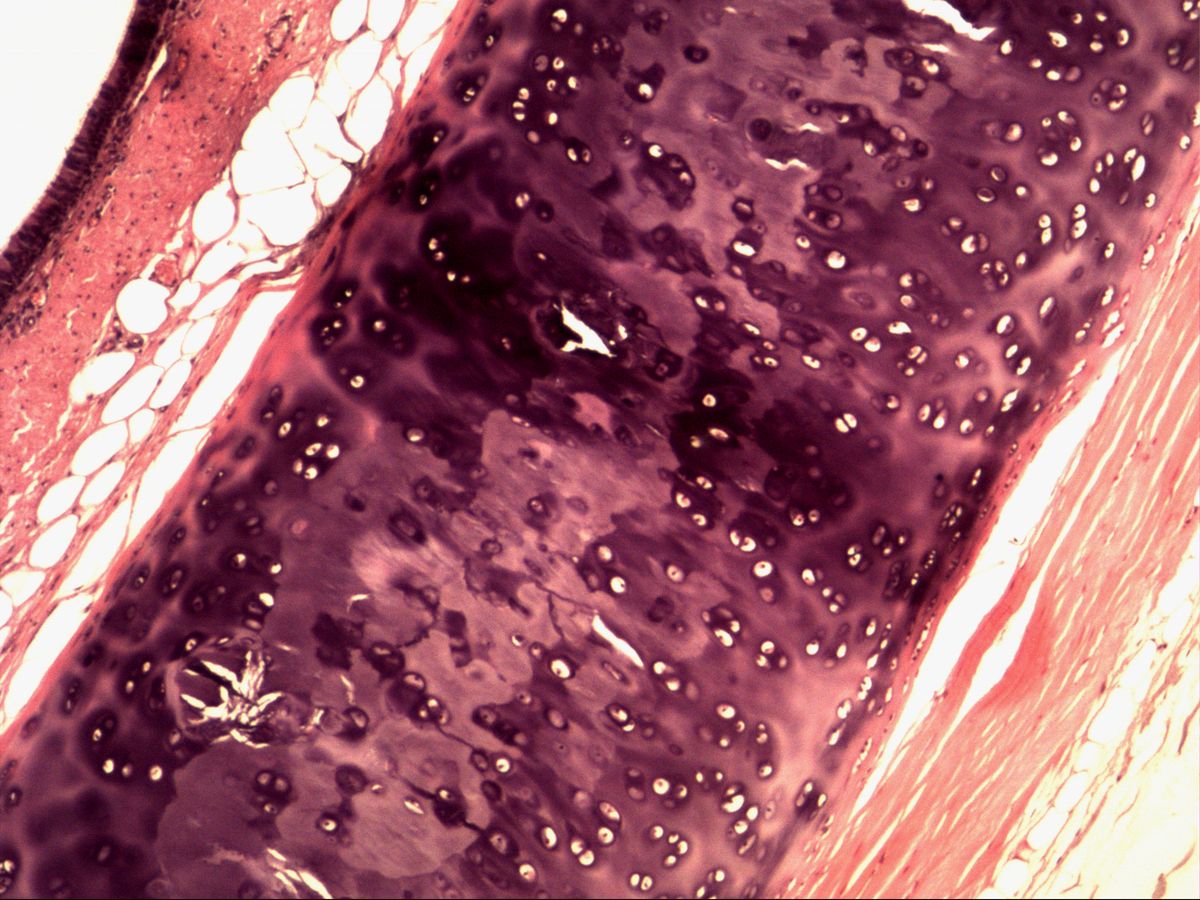
Labelled structures
This image has no labels – use the caption instead.
Hyaline cartilage is the most common type of cartilage. Its matrix looks homogeneous and glassy (Greek hyalos = glass) because the type II collagen fibrils have the same refractive index as the ground substance. The cartilage rings of the trachea show all typical components: chondrons, territorial matrix and the enveloping perichondrium.
How to recognise it
- Homogeneous, glassy, basophilic matrix without visible fibres
- Chondrocytes in lacunae, often in isogenous groups (2–8 cells)
- Dark territorial matrix around each chondron, paler interterritorial matrix
- Perichondrium with flattened cells at the edge
- No blood vessels within the cartilage
Structures to find
- Chondrocyte in lacuna
- Chondron (isogenous group, territory)
- Cartilage capsule
- Territorial matrix
- Interterritorial matrix
- Perichondrium
Structure
Chondrocytes lie in lacunae within the matrix. Cell division produces groups of 2–8 cells, the isogenous groups. Such a group together with the immediately surrounding, strongly basophilic matrix forms the chondron (territory). Directly around each lacuna lies the cartilage capsule, around that the territorial matrix. Between chondrons is the paler interterritorial matrix.
Externally the perichondrium surrounds the cartilage: an outer fibrous layer of dense connective tissue and an inner chondrogenic layer with progenitor cells. Cartilage itself is avascular and aneural.
Comparison: elastic cartilage and fibrocartilage
- Elastic cartilage (auricle, epiglottis): matrix additionally contains a dense network of elastic fibres (dark with elastic or resorcin–fuchsin stains), small chondrons of usually 1–3 cells, perichondrium present, does not calcify.
- Fibrocartilage (intervertebral disc, pubic symphysis, menisci): abundant type I collagen bundles running parallel or interwoven; chondrocytes single or in short rows between the bundles; usually no perichondrium.
- Hyaline cartilage: glassy matrix without visible fibres, large isogenous groups, perichondrium (except articular cartilage).
Microscopy tips
At low power the cartilage ring is a large violet, basophilic field between mucosa and adventitia. Move to the edge: there the chondrocytes become flat and spindle-shaped and merge into the perichondrium – a useful exam feature. In the centre large, rounded cells lie in isogenous groups with dark territorial matrix. Shrinkage often makes the cells retract from the lacunar wall; the white gap is an artefact. Differential from bone: bone is eosinophilic, lamellar and has vascular canals.
Function and clinical relevance
Hyaline cartilage resists compression: the strongly negatively charged proteoglycans (aggrecan) bind water that is squeezed out under load. It forms articular surfaces, costal cartilage, nasal, laryngeal and tracheal cartilage, and the embryonic skeletal model. Because it is avascular and nourished by diffusion it barely regenerates – cartilage damage in osteoarthritis is therefore largely irreversible. With age hyaline cartilage increasingly calcifies, e.g. costal and laryngeal cartilage.
Slide images
Test your knowledge
Assign the correct number in the image to each term.
Tap the requested structure in the image.
Multiple-choice questions on structure and function.
Frequently asked questions
Why is the cartilage matrix basophilic in H&E?
The proteoglycans contain many sulphated glycosaminoglycans (chondroitin and keratan sulphate) with negative charges that bind haematoxylin. Their concentration is highest around the cells, hence the dark territorial matrix.
What is the difference between a chondron and an isogenous group?
The isogenous group refers to the chondrocytes derived from a single cell. The chondron (territory) comprises this group together with its capsule and surrounding territorial matrix.
Does all hyaline cartilage have a perichondrium?
No. Articular cartilage has no perichondrium and is nourished by synovial fluid. Fibrocartilage usually lacks one as well.
Related topics
Updated: