Endochondral ossification
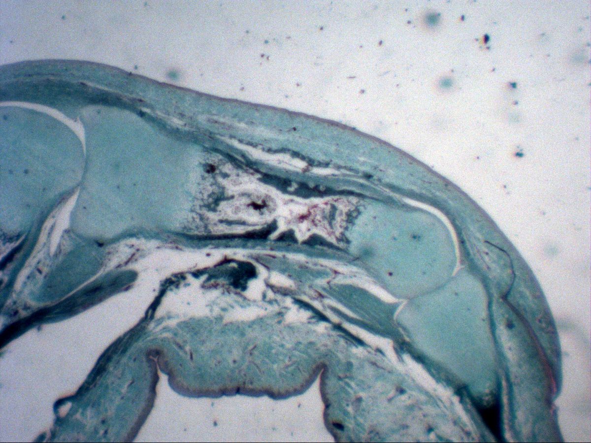
Fetal phalanx · trichrome
Labelled structures
This image has no labels – use the caption instead.
In endochondral (indirect) ossification a model of hyaline cartilage is laid down first and then gradually replaced by bone. Almost all bones of the axial and limb skeleton develop this way. The fetal phalanx shows all zones within a small area – from the resting cartilage of the epiphysis to the primary marrow cavity of the diaphysis.
How to recognise it
- Cartilage model with transition from resting cartilage to bone
- Columnar cartilage: cells in longitudinal rows
- Hypertrophic cartilage: large, vesicular chondrocytes with calcified septa
- Zone of erosion with capillaries, red blood cells and mesenchyme
- Perichondral bone collar beneath the periosteum
- Primary trabeculae with a cartilage core
Structures to find
- Epiphysis (cartilaginous) and diaphysis
- Reserve zone (hyaline cartilage)
- Columnar cartilage
- Hypertrophic (vesicular) cartilage
- Zone of erosion
- Primary marrow cavity
- Perichondral bone collar
- Periosteum and perichondrium
Process
First the perichondral bone collar forms around the middle of the diaphysis: the perichondrium becomes periosteum, whose osteoblasts lay down a bony sleeve around the cartilage by direct ossification. The enclosed cartilage hypertrophies and calcifies. Vessels with mesenchyme and osteoclasts (chondroclasts) then invade, resorb the cartilage and open the primary marrow cavity (primary ossification centre).
Osteoblasts deposit bone on the remaining calcified cartilage septa, forming primary trabeculae with a cartilage core. Ossification advances towards both epiphyses; later, secondary ossification centres appear there, and the epiphyseal plate remains between them as the growth zone.
Zones of the growth plate
- Reserve zone (resting hyaline cartilage): small, irregularly scattered chondrocytes.
- Proliferative zone (columnar cartilage): flattened cells that divide and stack into longitudinal columns.
- Hypertrophic zone (vesicular cartilage): greatly enlarged, bubble-like chondrocytes; the matrix between the columns calcifies.
- Zone of erosion (opening zone): capillaries and chondroclasts open the cartilage lacunae from the diaphyseal side; the chondrocytes die.
- Zone of ossification: osteoblasts deposit bone on the calcified longitudinal septa.
Microscopy tips
In the trichrome stain used here, cartilage matrix appears pale turquoise, bone and calcified septa dark green to reddish-brown, and red blood cells bright red. Start at the cartilaginous epiphysis at low power and follow the long axis towards the diaphysis: cells first flatten and line up in columns, then become large and vesicular, until the lacunae are finally open and contain blood and mesenchyme. Laterally you will find the dark bone collar beneath the periosteum – it extends further towards the epiphysis than the endochondral ossification front.
Clinical relevance
In achondroplasia (activating mutation of the FGFR3 gene) proliferation in the columnar zone is inhibited – long bones remain short while the intramembranously formed skull grows normally. In rickets (vitamin D deficiency) the hypertrophic cartilage fails to calcify properly, the erosion zone is irregular and the growth plates widen. Fractures through the growth plate in children (Salter–Harris classification) can lead to growth disturbances.
Slide images
Test your knowledge
Assign the correct number in the image to each term.
Tap the requested structure in the image.
Multiple-choice questions on structure and function.
Frequently asked questions
Is the bone collar formed endochondrally or intramembranously?
Intramembranously: the periosteal osteoblasts form it directly without a cartilage stage. Endochondral ossification of a long bone therefore always involves both mechanisms side by side.
Why do hypertrophic chondrocytes die?
The surrounding matrix calcifies and impedes diffusion of nutrients; they are also actively driven into programmed cell death. Before that they secrete VEGF and thereby attract blood vessels.
When do the growth plates close?
At the end of puberty, controlled mainly by oestrogens. Bone then replaces the growth-plate cartilage and longitudinal growth stops; an epiphyseal scar often remains visible on X-ray.
Related topics
Updated: