Learning path · Histology
Germ cells & embryonic tissue
Spermatogenesis in the seminiferous tubule, follicle maturation in the ovary and early embryonic development with gastrulation and neurulation – the basics of the germ layers in microscopic images.
-
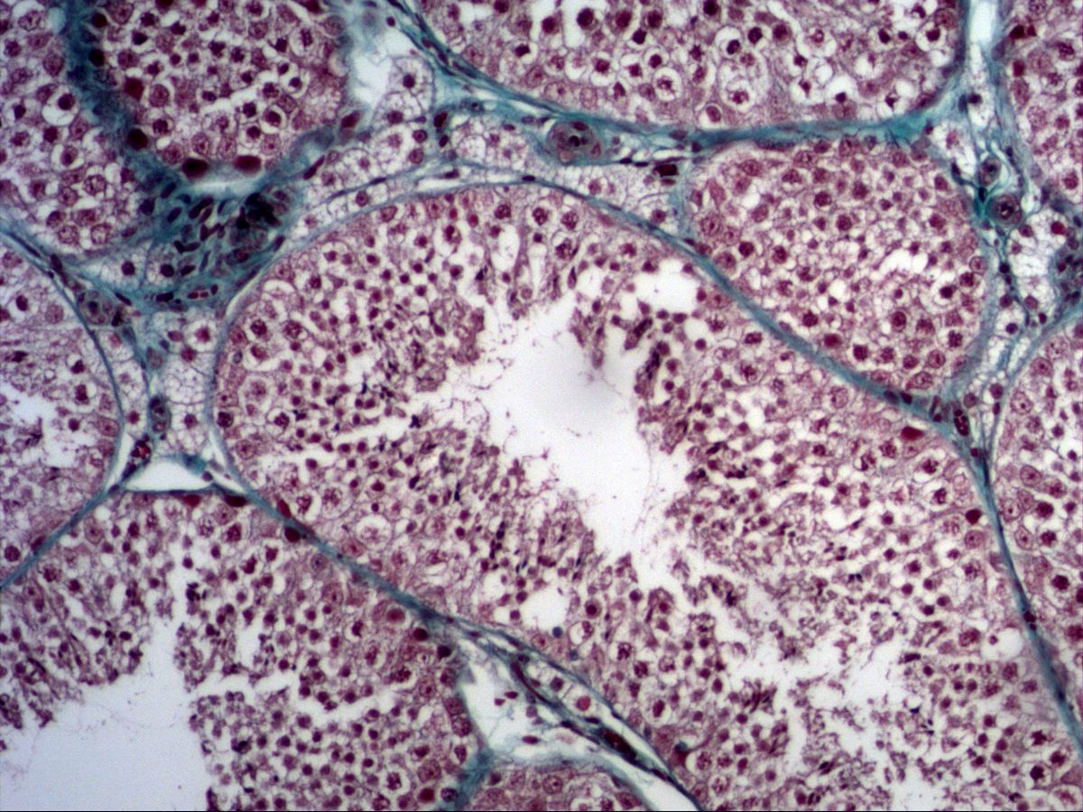
Spermatogenesis
Testis · trichrome
2 images 10 structures -
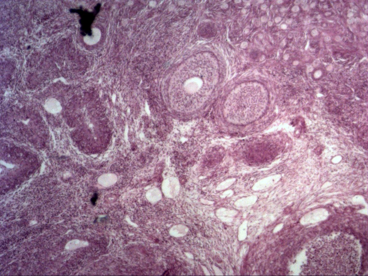
Oocytes and follicle maturation
Ovary · H&E
3 images 15 structures -
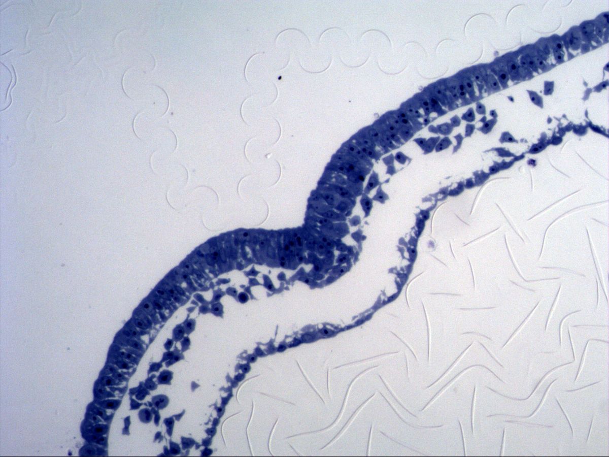
Gastrulation – early germ layers
Primitive streak · semithin section
2 images 8 structures -
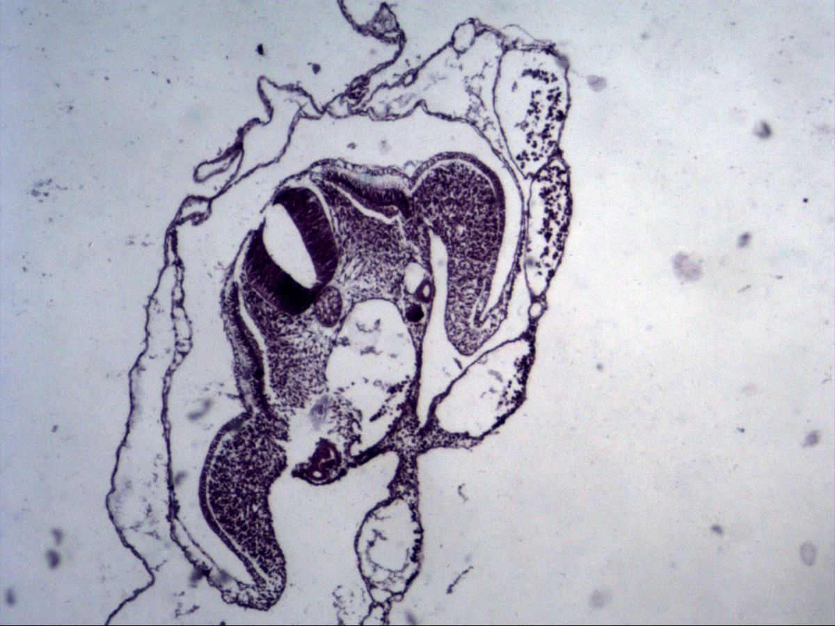
Neurulation – advanced germ layers
Chick embryo · H&E
2 images 10 structures
Test your knowledge
Mixed exam simulation: identify slides, name highlighted structures and answer knowledge questions. Choose your areas and off you go.