Neurulation – advanced germ layers
Chick embryo · H&E
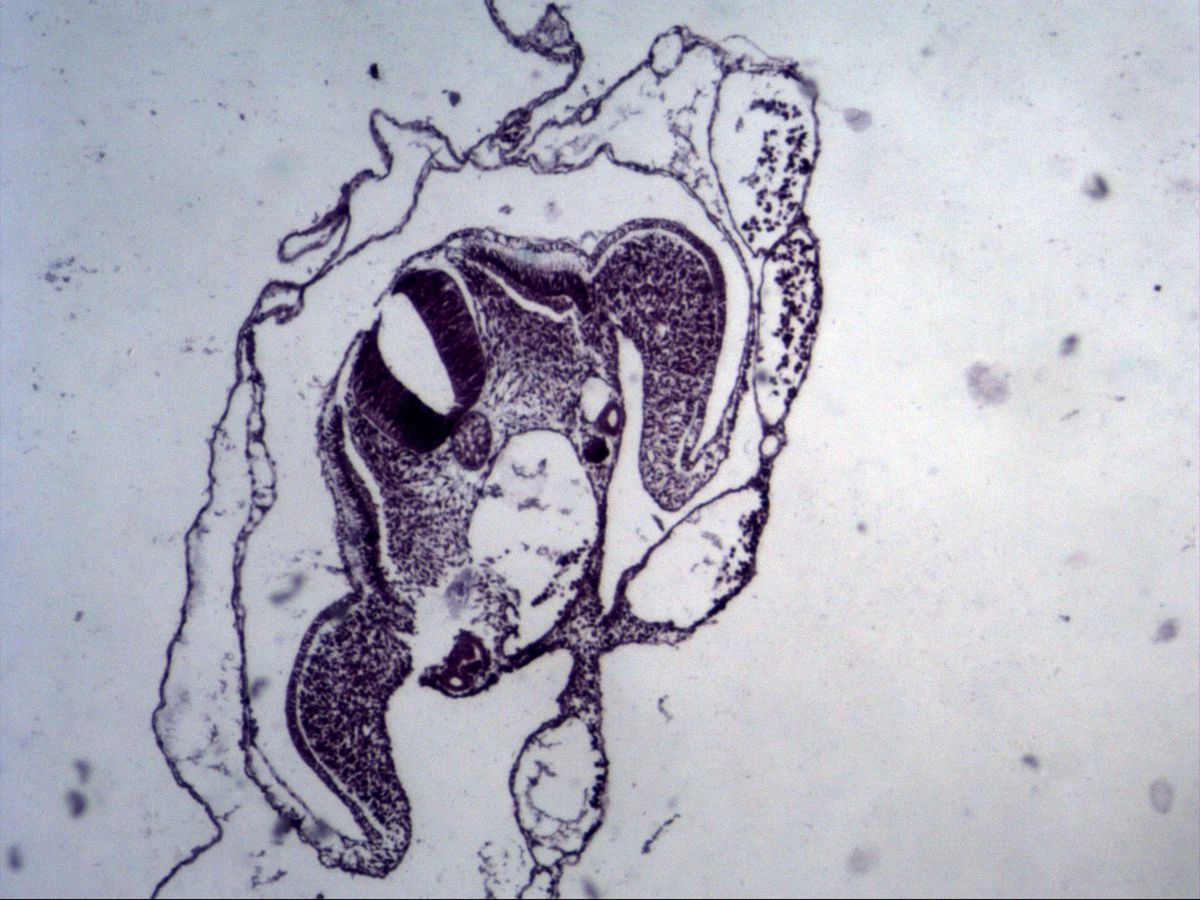
Labelled structures
This image has no labels – use the caption instead.
During neurulation the neural plate forms from the ectoderm above the notochord. It folds up into neural folds, which fuse in the midline to form the neural tube – the primordium of the brain and spinal cord. At the same time the mesoderm on each side divides into paraxial, intermediate and lateral plate mesoderm, and the embryo folds into a cylindrical body.
The cross-section through a chick embryo (H&E) shows this advanced organisation of the germ layers: the neural tube lies dorsally, the notochord beneath it, the mesodermal divisions laterally, all surrounded by ectoderm and the extraembryonic membranes.
How to recognise it
- Thick-walled neural tube with a slit-like lumen
- Round, vacuolated notochord ventral to the neural tube
- Loose mesenchyme of the paraxial mesoderm on both sides
- Lateral plate mesoderm with coelomic cleft, ectoderm outside
- Thin extraembryonic membranes around the embryo
Structures to find
- Neural tube
- Notochord
- Paraxial mesoderm (sclerotome, myotome, dermatome)
- Intermediate mesoderm (Wolffian duct)
- Lateral plate mesoderm
- Coelom
- Ectoderm
- Endoderm
Structure
The neural tube consists of a thick, pseudostratified neuroepithelium with crowded nuclei around a slit-like lumen (the future central canal or ventricle). Ventral to it lies the notochord, a round rod of large, vacuolated cells. Lateral to the neural tube is the paraxial mesoderm, which segments into somites (sclerotome, myotome, dermatome). Further laterally is the narrow intermediate mesoderm, which gives rise to the mesonephros and Wolffian duct. Outermost lies the lateral plate mesoderm, which splits into a somatic and a splanchnic layer; the space between is the intraembryonic coelom. Ectoderm covers the embryo on the outside, and endoderm lines the gut tube.
Function
The notochord is an important signalling centre: among other factors it secretes Sonic hedgehog and thereby induces the neural plate, the ventral floor plate of the neural tube and the sclerotome. The neural tube gives rise to the CNS and retina, the neural folds to the neural crest (spinal ganglia, autonomic ganglia, melanocytes, adrenal medulla). The sclerotome forms the vertebrae, the myotome skeletal muscle and the dermatome the dermis of the back. The somatic layer of the lateral plate forms the body wall and limbs, the splanchnic layer the gut wall musculature and heart. The coelom gives rise to the body cavities.
Microscopy tips
At low magnification first look for the neural tube: the most conspicuous, darkest structure with a thick epithelium and narrow lumen. Directly ventral to it you will find the small, round notochord with pale vacuoles – it defines the midline and the orientation of the section. The specimen is often embedded obliquely or rotated, so always orient yourself by the neural tube and notochord, not by the edge of the image. Laterally follow loose mesenchyme and the mesodermal plates, and on the outside thin extraembryonic membranes (amnion, chorion). Blood vessels can be recognised by nucleated erythrocytes, which in birds remain nucleated even in adults.
Clinical relevance
If the neural tube does not close completely, neural tube defects result: caudally spina bifida (up to myelomeningocele), cranially anencephaly. In humans closure occurs as early as the 4th embryonic week, often before the pregnancy is noticed. Folic acid supplementation before conception markedly reduces the risk. Raised alpha-fetoprotein in maternal serum or amniotic fluid may indicate an open defect; notochordal remnants can rarely give rise to a chordoma.
Slide images
Test your knowledge
Assign the correct number in the image to each term.
Tap the requested structure in the image.
Multiple-choice questions on structure and function.
Frequently asked questions
How do I recognise the notochord?
It is a small, round, sharply delimited rod of cells directly ventral to the neural tube. Its cells contain pale vacuoles.
What does the intermediate mesoderm become?
It forms the urogenital system: pronephros, mesonephros with Wolffian duct, metanephros and the gonadal ridge.
Why is a chick embryo used?
Chick embryos are readily available, develop outside the mother and show neurulation in a very similar way to mammals.
Related topics
Updated: