Mucous (gelatinous) connective tissue
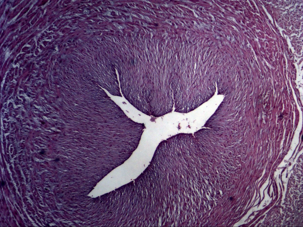
Umbilical cord · H&E
Labelled structures
This image has no labels – use the caption instead.
Mucous (gelatinous) connective tissue is an embryonic connective tissue that is practically absent in adults. Its classic location is the umbilical cord, where it embeds the three umbilical vessels as Wharton's jelly and protects them from kinking and compression.
How to recognise it
- Abundant pale, homogeneous ground substance, few fine fibres
- Stellate or spindle-shaped branched fibroblasts connected by processes
- No free cells, hardly any capillaries
- In the umbilical cord: two arteries with thick muscle wall, one vein
- Single-layered amniotic epithelium on the surface
Structures to find
- Gelatinous ground substance (Wharton's jelly)
- Stellate fibroblasts
- Umbilical artery with smooth muscle
- Umbilical vein
- Amniotic epithelium
Structure
The tissue consists mainly of a water-rich, gelatinous ground substance rich in hyaluronic acid. Within it lie loosely scattered, stellate or spindle-shaped branched fibroblasts (mesenchyme-like cells) whose processes contact one another. Collagen fibres are initially sparse and fine but increase as pregnancy advances.
The umbilical cord contains two umbilical arteries with a thick smooth-muscle wall and a narrow, often star-shaped lumen, and one umbilical vein with a wider lumen. Externally it is covered by a single-layered amniotic epithelium. Vasa vasorum and nerves are absent.
Function
Hyaluronic acid binds large amounts of water and makes Wharton's jelly turgid and elastic. It acts as a cushion that protects the umbilical vessels from compression, torsion and knotting, thereby securing blood flow between placenta and fetus. The cells of Wharton's jelly are multipotent mesenchymal stromal cells that can be differentiated experimentally into cartilage, bone or fat cells, which is why they are of interest in stem cell research.
Microscopy tips
At low power orient yourself by the vessels: the umbilical artery stands out with its thick, circular muscle wall and collapsed lumen. Between the vessels lies the pale, homogeneous jelly. At high power you can see the branched fibroblasts distributed like a loose network in the pale ground substance; their nuclei are elongated to oval. The pale gaps are ground substance, which barely stains in paraffin sections. Differential: loose connective tissue contains many more fibres, vessels and free cells; mesenchyme is even more cellular and fibre-free.
Clinical relevance
A single umbilical artery (one instead of two arteries) occurs in about 1 % of pregnancies and is associated with malformations, e.g. of the kidneys or heart; the number of vessels is therefore checked on ultrasound and after birth. A thin cord with little jelly is more susceptible to compression. Cord blood is used to measure pH and blood gases and can be stored as a source of stem cells.
Slide images
Test your knowledge
Assign the correct number in the image to each term.
Tap the requested structure in the image.
Multiple-choice questions on structure and function.
Frequently asked questions
What is Wharton's jelly?
This is the name for the mucous connective tissue of the umbilical cord. It consists of ground substance rich in hyaluronic acid with branched fibroblasts and surrounds the umbilical vessels.
How do umbilical artery and vein differ histologically?
The arteries have a thicker, strong muscle wall and a narrow lumen that is often contracted into a star shape. The vein has a thinner wall and a wider lumen.
Where is mucous connective tissue found in adults?
Practically nowhere; at most the pulp of young teeth is similarly built. It is characteristic of the embryo and the umbilical cord.
Related topics
Updated: