Fibroblasts and fibrocytes
Cell culture · Giemsa
Labelled structures
This image has no labels – use the caption instead.
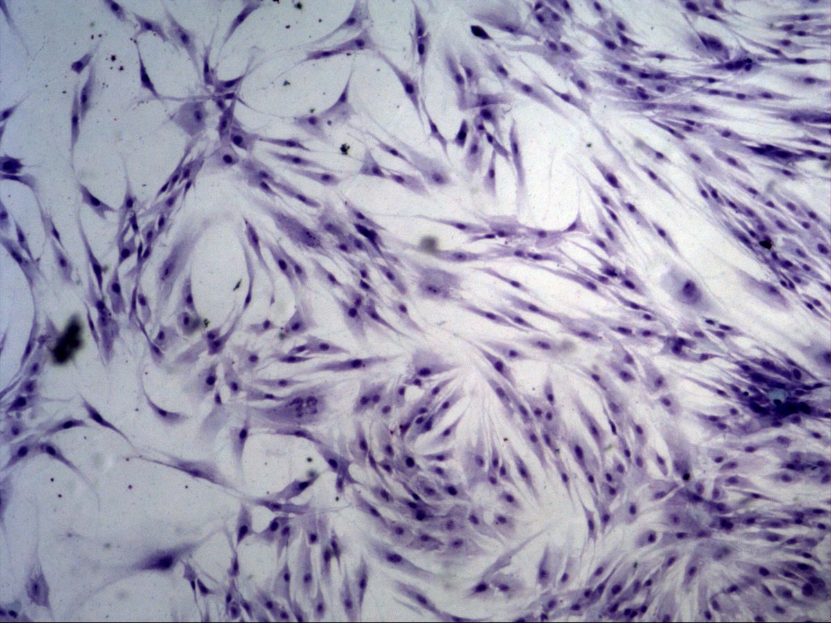
Fibroblasts are the principal resident cells of connective tissue. In tissue sections usually only their slender nuclei are visible between the fibres. In cell culture, by contrast, they grow flattened on the slide, so Giemsa staining reveals the entire cell shape with nucleus, cytoplasm and processes.
How to recognise it
- Spindle-shaped to stellate, flattened cells with long processes
- Large, oval, pale nucleus with prominent nucleoli (active fibroblast)
- Pale violet, slightly basophilic cytoplasm
- Fibrocyte: smaller, slender, darker nucleus, little cytoplasm
- Parallel or whorled arrangement at high cell density
Structures to find
- Fibroblast with nucleus and nucleolus
- Cytoplasmic processes
- Fibrocyte (slender resting form)
- Borders of neighbouring cells
Structure
The active fibroblast is a large, spindle-shaped to stellate cell with several processes. Its nucleus is large, oval and pale (euchromatic) with prominent nucleoli; the cytoplasm is basophilic owing to abundant rough ER. These features indicate high protein synthesis.
The fibrocyte is the resting form: smaller and slender, with a dark, flattened, heterochromatin-rich nucleus and little cytoplasm. Both forms are functional states of the same cell lineage and can convert into each other, e.g. during wound healing.
Function
Fibroblasts produce the entire extracellular matrix of connective tissue: procollagen (which polymerises into collagen fibrils outside the cell), elastin, fibrillin, proteoglycans and glycoproteins such as fibronectin. They also degrade matrix via metalloproteinases, keeping the tissue in balance. During wound healing they migrate in, proliferate and partly become myofibroblasts, which contract the wound edges using actin filaments. Fibroblasts also release growth factors and cytokines, thereby influencing inflammation and angiogenesis.
Microscopy tips
In Giemsa-stained culture nuclei appear dark violet and cytoplasm pale violet. At low power you see a dense network of spindle-shaped cells, which line up in parallel or in whorls at high density. At high power look for oval, pale nuclei with 1–3 nucleoli and long, thin tapering processes. Note the difference from tissue sections: there fibrocytes are recognisable only as slender dark nuclei between collagen fibres, without visible cell borders.
Clinical relevance
Excessive fibroblast activity leads to fibrosis, for example in the lung, liver (cirrhosis) or skin (scleroderma), and to hypertrophic scars and keloids. In vitamin C deficiency (scurvy) fibroblasts cannot hydroxylate proline and lysine in collagen; unstable collagen causes bleeding gums and poor wound healing. Fibroblast cultures from skin biopsies are used to diagnose genetic metabolic and collagen disorders, for example suspected osteogenesis imperfecta or lysosomal storage diseases.
Slide images
Test your knowledge
Assign the correct number in the image to each term.
Tap the requested structure in the image.
Multiple-choice questions on structure and function.
Frequently asked questions
Why are fibroblasts studied in cell culture?
In culture the cells grow flat in a monolayer, so their entire shape including processes becomes visible. In tissue sections they can hardly be distinguished from the surrounding fibres.
What does Giemsa staining show?
Giemsa is an azure–eosin–methylene blue stain. Nuclear chromatin stains dark violet and basophilic cytoplasm blue-violet, so nuclei, nucleoli and cell outlines are clearly visible.
Are fibroblasts and fibrocytes different cell types?
No, they are two activity states of the same cell. The fibroblast is synthetically active, the fibrocyte is resting but can be reactivated.
Related topics
Updated: