Adipose tissue
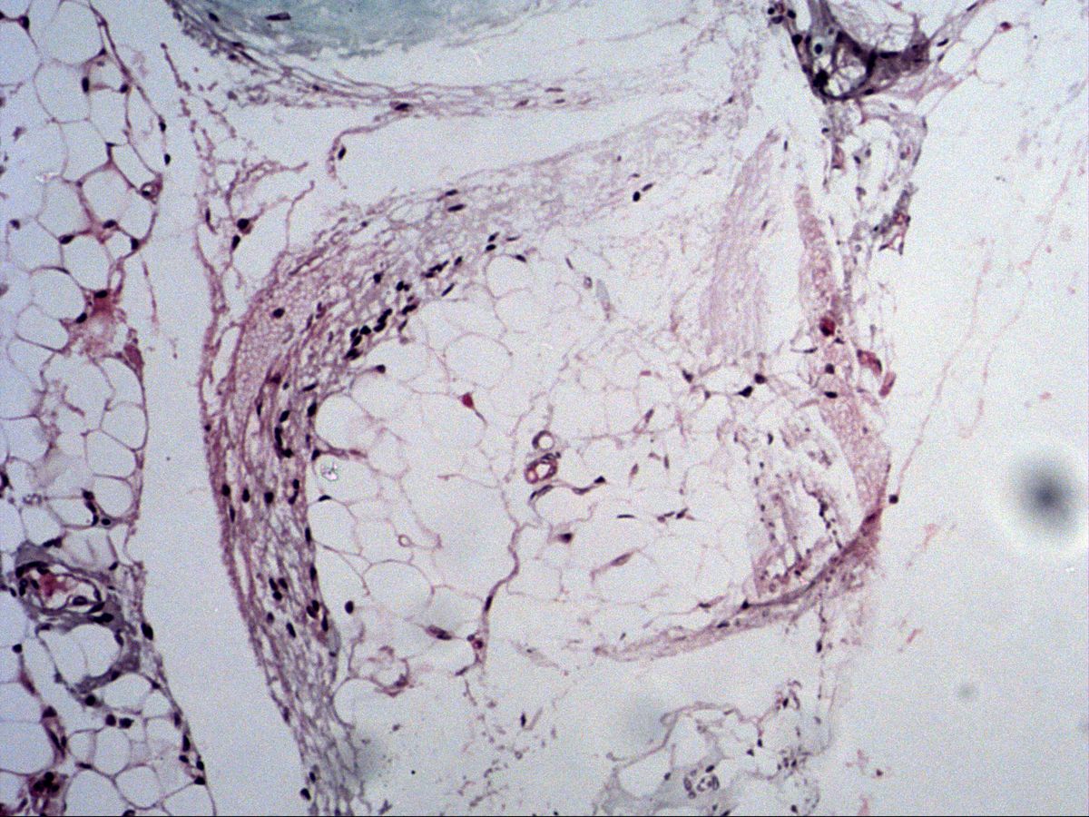
Neurovascular bundle · trichrome
Labelled structures
This image has no labels – use the caption instead.
Adipose tissue is a specialised connective tissue of tightly packed adipocytes surrounded by fine connective tissue septa and a dense capillary network. In routine sections the fat is dissolved by alcohol and xylene, so the cells appear as empty, honeycomb-like spaces. The section shown here comes from the connective tissue of a neurovascular bundle.
How to recognise it
- Large, empty, polygonal cell spaces (fat dissolved)
- Honeycomb pattern with thin membrane rims
- Flattened, peripheral nucleus: signet-ring cell
- Fine connective tissue septa with capillaries
- Multilocular cells (brown fat): many small vacuoles, central nucleus
Structures to find
- Unilocular adipocytes
- Signet-ring, flattened nucleus
- Connective tissue septa
- Capillaries and small vessels
- For comparison: multilocular adipocytes (brown fat, textbook)
Structure
White (unilocular) adipose tissue: each cell contains a single large lipid droplet that pushes the nucleus and cytoplasm to the periphery as a thin rim – in section this produces the signet-ring cell appearance. Each adipocyte is surrounded by a basal lamina and reticular fibres; groups of cells form lobules separated by septa.
Brown (multilocular) adipose tissue: smaller cells with many small lipid droplets, a central round nucleus and abundant mitochondria; the tissue is very rich in capillaries and nerves.
Function and occurrence
White adipose tissue serves as storage fat (energy reserve, mainly subcutis, omentum, mesentery), as structural fat (mechanical cushion, e.g. sole of the foot, buccal fat pad, renal bed) and as thermal insulation. It is also an endocrine organ: adipocytes secrete leptin and adiponectin.
Brown adipose tissue produces heat via the uncoupling protein UCP-1 (thermogenin) (non-shivering thermogenesis). It is abundant in newborns (between the shoulder blades, in the neck, around the kidneys) and persists only in remnants in adults.
Microscopy tips
At low power adipose tissue is recognised by a fine, pale honeycomb pattern of large, empty, polygonal spaces. At high power look for a nucleus flattened against the edge of the vacuole – the signet-ring shape. Many nuclei between the cells, however, belong to capillary endothelial cells or fibrocytes of the septa.
Pitfall: mucus-secreting cells or cartilage with shrinkage artefacts may look similarly empty. Demonstrating fat requires frozen sections stained with Sudan or Oil Red O.
Clinical relevance
In obesity adipocytes mainly increase in size (hypertrophy); enlarged cells release more inflammatory mediators, which is linked to insulin resistance. The lipoma is the most common benign soft-tissue tumour and consists of mature white adipose tissue, often with a thin capsule. The rare hibernoma is a benign tumour of brown fat. Its malignant counterpart is liposarcoma, in which atypical lipoblasts with scalloped nuclei are diagnostically important. With marked weight loss adipocytes shrink, but their number remains largely unchanged.
Slide images
Test your knowledge
Assign the correct number in the image to each term.
Tap the requested structure in the image.
Multiple-choice questions on structure and function.
Frequently asked questions
Why are fat cells empty in sections?
During paraffin embedding, alcohol and xylene dissolve the lipids. What remains are the thin rims of cytoplasm, the cell membranes and the flattened nucleus.
What do unilocular and multilocular mean?
Unilocular adipocytes (white fat) contain a single large lipid droplet. Multilocular adipocytes (brown fat) contain many small droplets and numerous mitochondria.
Is adipose tissue a connective tissue?
Yes, it is classed as a specialised connective tissue because adipocytes derive from mesenchymal precursor cells. It is, however, cell-rich and poor in matrix.
Related topics
Updated: