Learning path · Histology
Epithelial tissue
Epithelia cover outer and inner surfaces. Learn to classify them reliably by layering (simple, pseudostratified, stratified), cell shape (squamous, cuboidal, columnar) and apical specialisations such as microvilli, cilia and stereocilia.
-
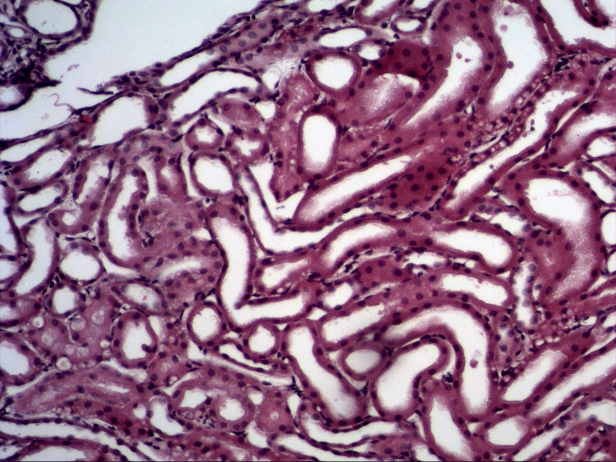
Simple squamous and simple cuboidal epithelium
Kidney · H&E
3 images 10 structures -
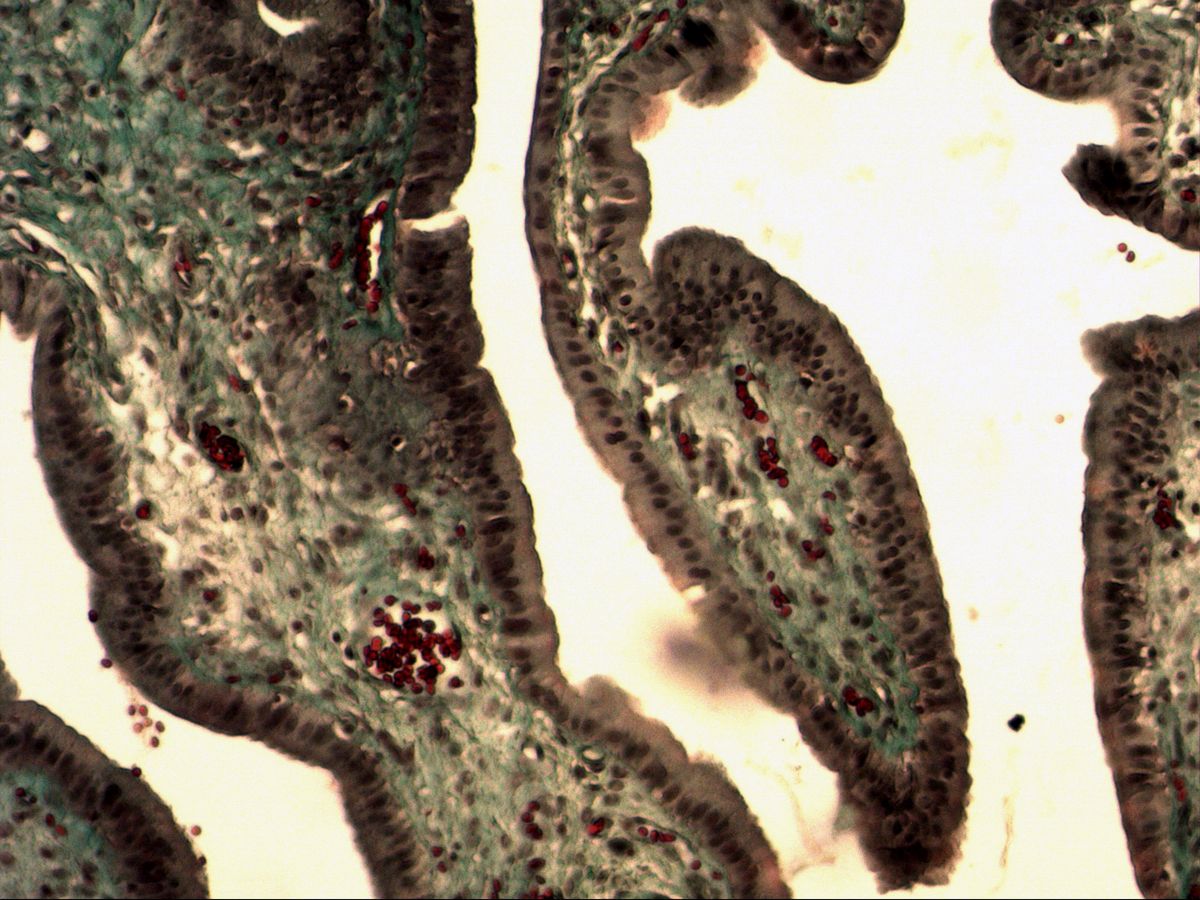
Simple columnar epithelium
Gallbladder · trichrome
3 images 14 structures -
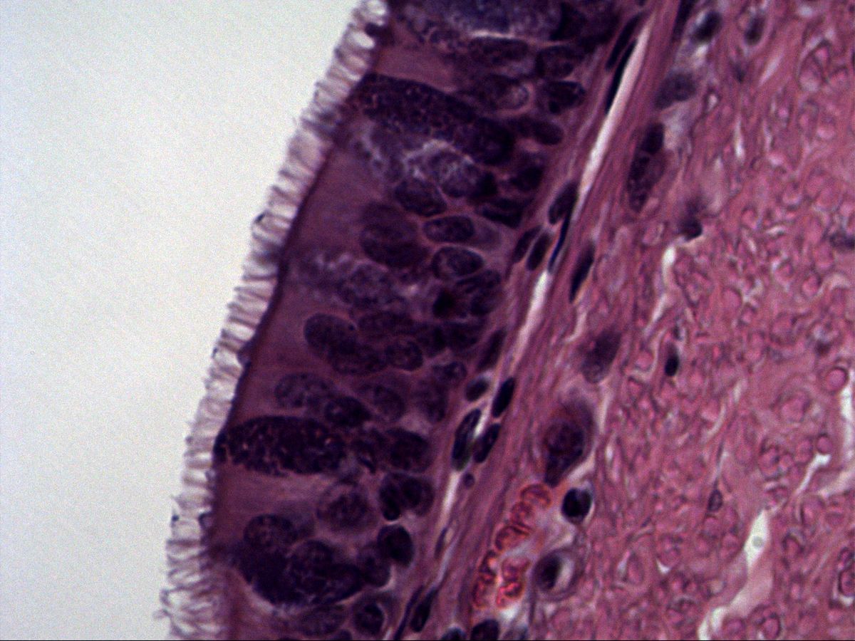
Pseudostratified columnar ciliated epithelium
Trachea · H&E
1 images 7 structures -
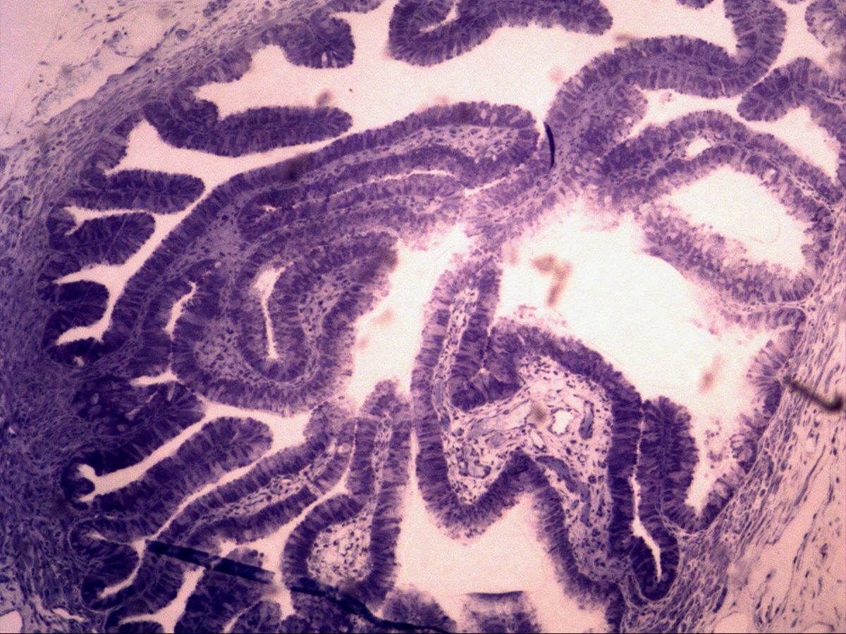
Ciliated epithelium of the uterine tube
Uterine tube · semithin section, toluidine blue
3 images 10 structures -
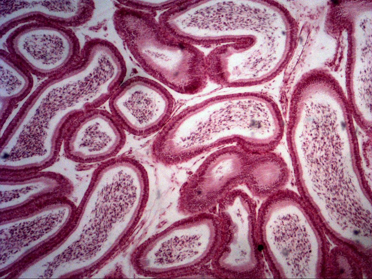
Pseudostratified columnar epithelium with stereocilia
Epididymis · H&E
4 images 13 structures -
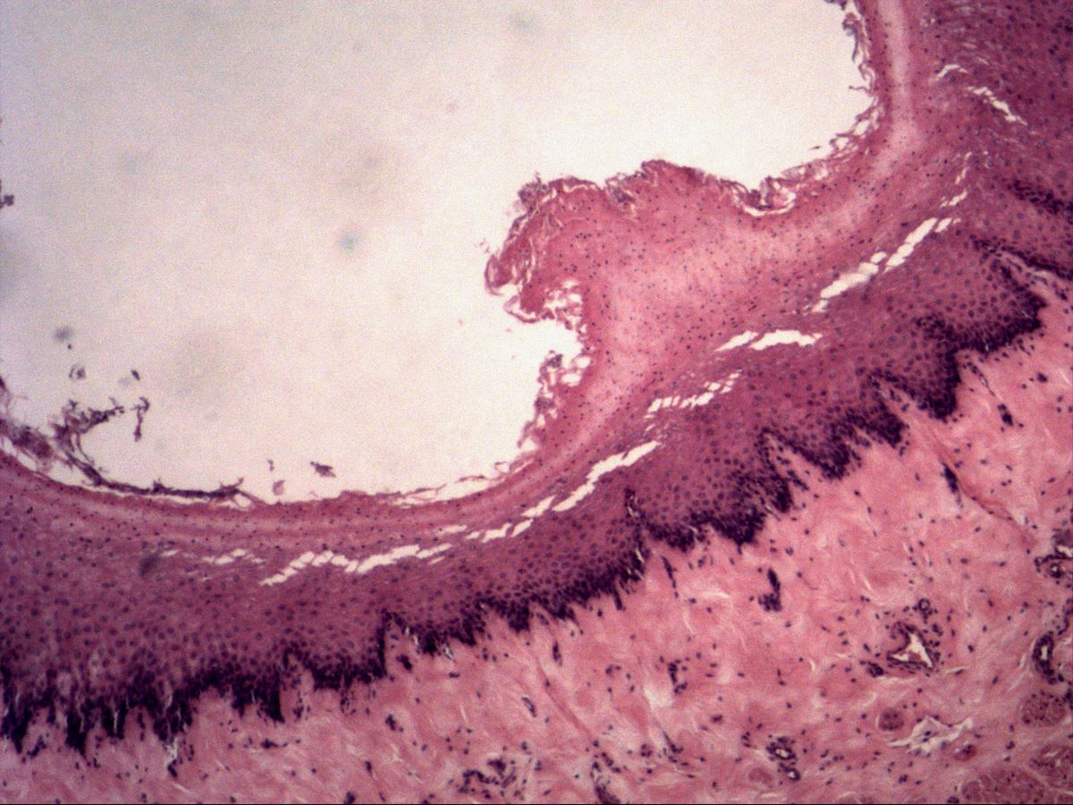
Non-keratinised stratified squamous epithelium
Oesophagus · H&E
6 images 15 structures -
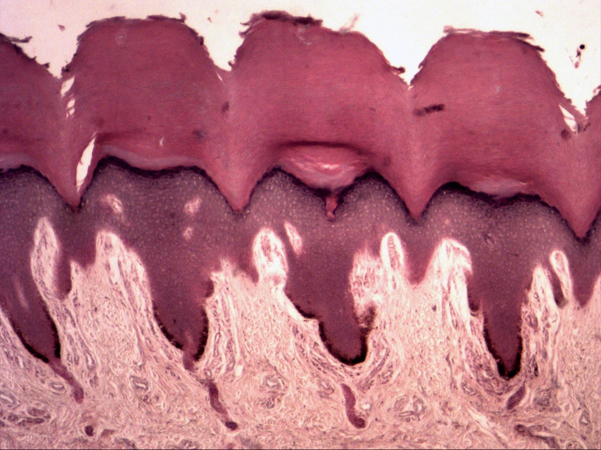
Keratinised stratified squamous epithelium
Fingertip skin · H&E
4 images 14 structures -
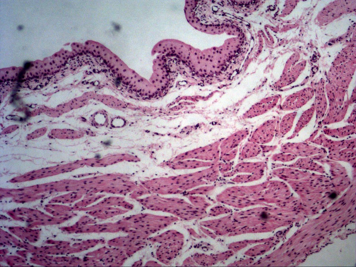
Urothelium (transitional epithelium)
Urinary bladder · H&E
1 images 6 structures
Test your knowledge
Mixed exam simulation: identify slides, name highlighted structures and answer knowledge questions. Choose your areas and off you go.