Non-keratinised stratified squamous epithelium
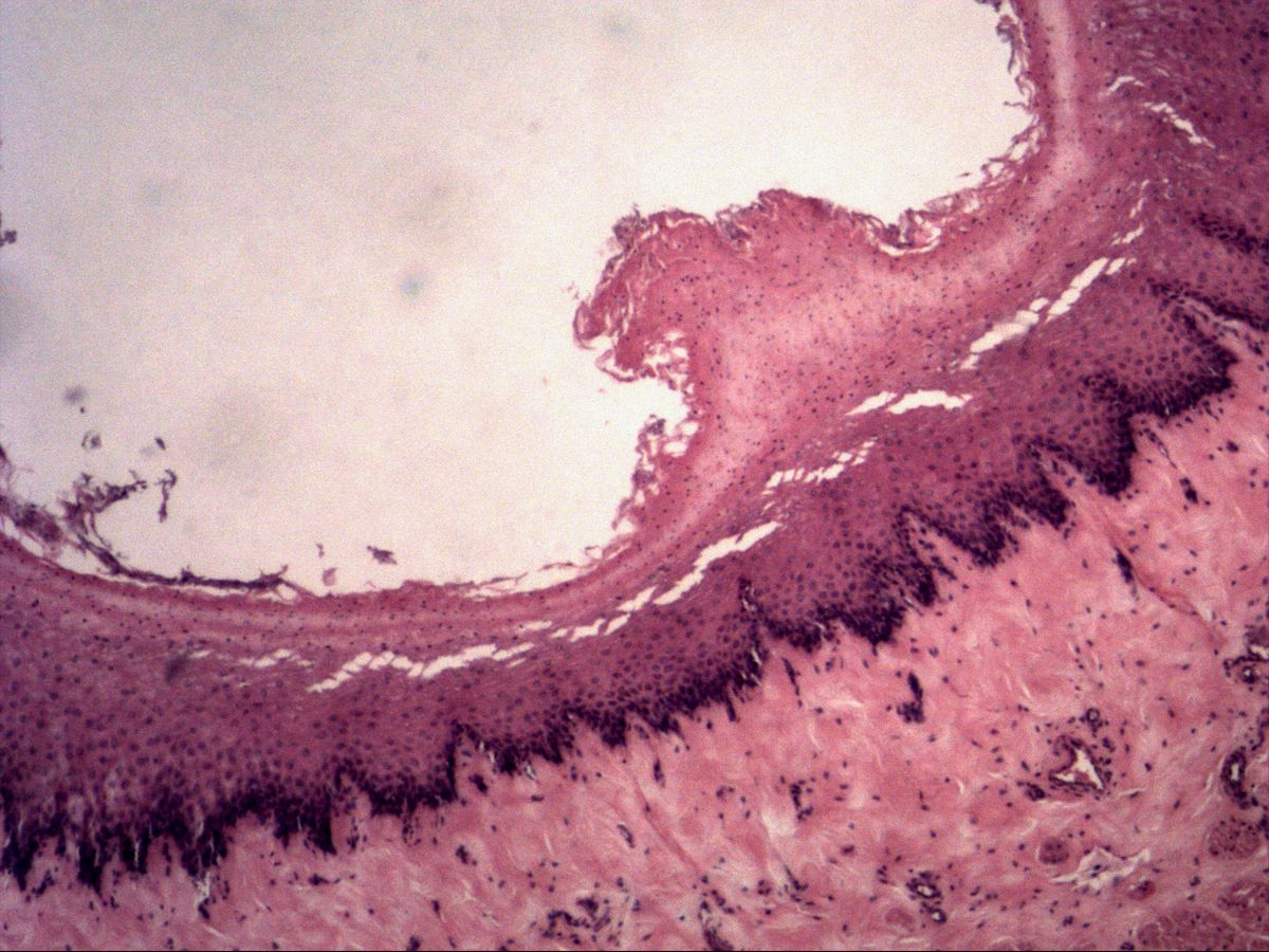
Oesophagus · H&E
Labelled structures
This image has no labels – use the caption instead.
Non-keratinised stratified squamous epithelium is typical of mechanically stressed zones. It occurs in the oesophagus and oral cavity as well as in the anal canal and the vagina, and also in the cornea and conjunctiva of the eye, and in the terminal part of the male urethra. Three layers can be recognised: stratum basale, stratum intermedium and stratum superficiale (with bulging cells).
In a stratified epithelium only the lowest cell layer rests on the basement membrane. From basal to apical the cells become progressively flatter – the shape of the uppermost cells gives the epithelium its name. 'Non-keratinised' means that even the surface cells still have a nucleus and do not form an anuclear horny layer. Connective tissue papillae reach deep into the epithelium and interlock it with the lamina propria.
How to recognise it
- Many cell layers, only the lowest on the basement membrane
- Cells become progressively flatter towards the surface
- Nuclei present up to the uppermost layer
- No anuclear horny layer
- Wavy base due to connective tissue papillae
Structures to find
- Stratum basale
- Stratum intermedium
- Stratum superficiale with typical flat nuclei
- Basement membrane
- Connective tissue papilla
- Lamina propria
Structure
The stratum basale consists of small, densely packed, cuboidal to columnar cells with dark nuclei; this is where stem cells divide. Above it, the stratum intermedium (spinosum) contains large polygonal cells with round, pale nuclei, connected by numerous desmosomes. In the stratum superficiale the cells become flat and their nuclei flattened and pyknotic, but still present. The cells contain abundant glycogen and therefore look pale in H&E. Tall connective tissue papillae with capillaries supply the avascular epithelium.
Function
Many cell layers protect against mechanical stress, for example from a food bolus: surface cells are constantly shed and replaced from below. Because there is no horny layer, the surface remains moist and pliable; it is kept wet by saliva or glandular secretions. Desmosomes anchor the cells firmly to one another and distribute tensile forces. Unlike the epidermis, its barrier against water loss is weak – which is unnecessary on moist surfaces. The epithelium renews itself quickly, so small mucosal injuries usually heal fast and without scarring.
Microscopy tips
At low magnification you see a thick epithelial band with a dark, wavy base – the waves are caused by the connective tissue papillae. At high magnification follow a column of cells from basal to apical and watch them become flatter. Decisive for 'non-keratinised': nuclei up to the surface, no anuclear pink horny layer and no stratum granulosum. Differential diagnosis from urothelium: there the umbrella cells are large, dome-shaped and often binucleate rather than flat. Further out in the oesophageal wall follow the submucosa with glands and the muscular layer.
Clinical relevance
With chronic reflux of gastric acid the squamous epithelium of the lower oesophagus can transform into a columnar epithelium with goblet cells (Barrett's oesophagus). This metaplasia is a precancerous condition for adenocarcinoma. Smoking and alcohol, by contrast, favour squamous cell carcinoma. In reflux oesophagitis the stratum basale becomes thicker and the papillae reach unusually close to the surface – signs of increased regeneration.
Slide images
Test your knowledge
Assign the correct number in the image to each term.
Tap the requested structure in the image.
Multiple-choice questions on structure and function.
Frequently asked questions
How can I tell that the epithelium is non-keratinised?
Even the most superficial cells still have a nucleus, and there is no anuclear, homogeneously pink horny layer and no stratum granulosum.
Why do the cells of the stratum spinosum look 'spiny'?
During tissue processing the cells shrink but remain attached at their desmosomes. These contact points then appear as fine spines between the cells.
Where does non-keratinised stratified squamous epithelium occur?
In the oral cavity, oesophagus, anal canal, vagina and ectocervix, on the cornea and conjunctiva of the eye, and in the terminal part of the male urethra.
Related topics
Updated: