Pseudostratified columnar epithelium with stereocilia
Epididymis · H&E
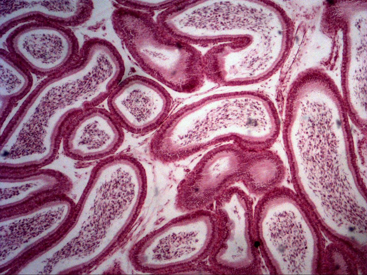
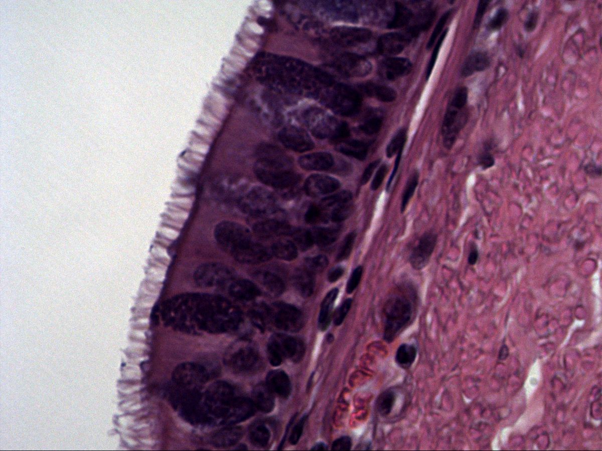
Labelled structures
This image has no labels – use the caption instead.
The two-row (pseudostratified) columnar epithelium has an apical surface specialisation: the stereocilia ('long microvilli') improve absorption thanks to the larger surface area.
The ductus epididymidis is a highly coiled duct several metres long, so a section shows many similar profiles side by side. Its epithelium is pseudostratified with two rows of nuclei: tall principal cells with elongated nuclei and long stereocilia, and small round basal cells at the basement membrane. The smooth, uniformly tall luminal contour and the sperm in the lumen make this slide easy to recognise.
How to recognise it
- Two rows of nuclei: elongated principal cell nuclei, round basal cell nuclei
- Long tufted stereocilia without a basal body line
- Smooth, uniformly tall luminal contour
- Sperm in the lumen
- Thin layer of smooth muscle cells around the duct
Structures to find
- Stereocilia
- Nuclei of principal cells
- Nuclei of basal cells
- Basement membrane
- Sperm in the lumen
- Smooth muscle cells
Structure
The principal cells extend from the basement membrane to the lumen; their elongated nucleus lies in the lower half of the cell and forms the upper row of nuclei. The basal cells are small, round to triangular and rest directly on the basement membrane – they form the lower row of nuclei. Apically the principal cells carry tufts of stereocilia: very long, partly branched microvilli (about 10 µm) with an actin filament core, without basal bodies and non-motile. Around the duct lies a thin layer of smooth muscle cells embedded in loose connective tissue.
Function
In the epididymis spermatozoa mature and are stored. The principal cells absorb most of the fluid coming from the testis – the stereocilia enlarge the surface for this – and take up residual bodies and cell debris. At the same time they secrete glycoproteins, glycerophosphocholine and sialic acid, which modify the sperm membrane. Only here do the sperm acquire the capacity for directed movement. The smooth muscle moves sperm towards the ductus deferens by peristaltic contractions.
Microscopy tips
At low magnification you see many round to oval duct profiles with a smooth inner contour and sperm in the lumen. At high magnification look for two rows of nuclei (elongated above, round below) and the tufts of clumped stereocilia. Differential diagnosis from the ductus deferens: same epithelium, but a very thick three-layered muscular wall and a star-shaped lumen. From the efferent ductules: there the luminal contour is 'saw-toothed' with groups of tall and short cells, and true motile cilia are present.
Slide images
Test your knowledge
Assign the correct number in the image to each term.
Tap the requested structure in the image.
Multiple-choice questions on structure and function.
Frequently asked questions
Are stereocilia true cilia?
No. Stereocilia are particularly long microvilli with an actin filament core. They have no microtubules, no basal bodies and are non-motile.
Why is the epithelium called two-row (pseudostratified)?
All cells rest on the basement membrane, but the nuclei of principal cells and basal cells lie at two different levels.
Where else do stereocilia occur?
In the ductus deferens and – in a modified, rigid form – on the hair cells of the inner ear, where they serve mechanotransduction.
Related topics
Updated: