Pseudostratified columnar ciliated epithelium
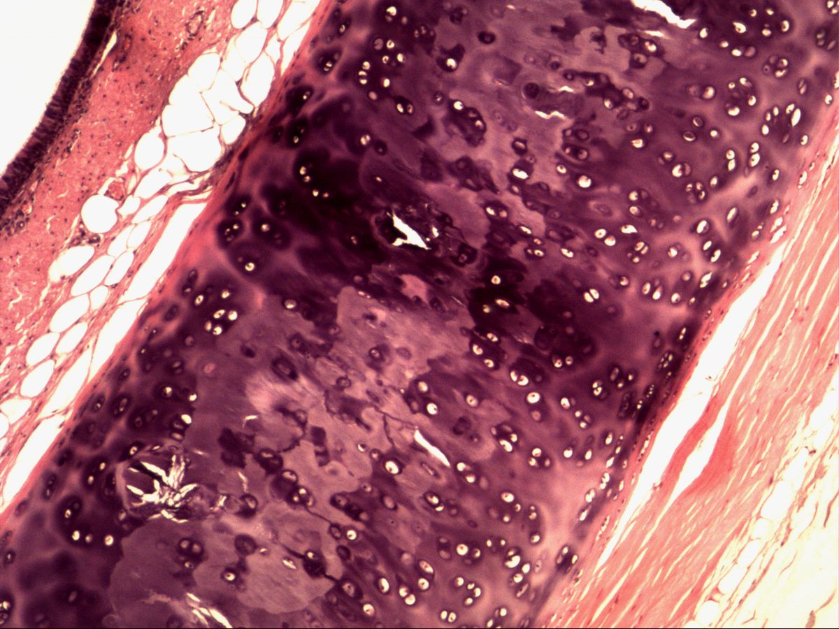
Trachea · H&E
Labelled structures
This image has no labels – use the caption instead.
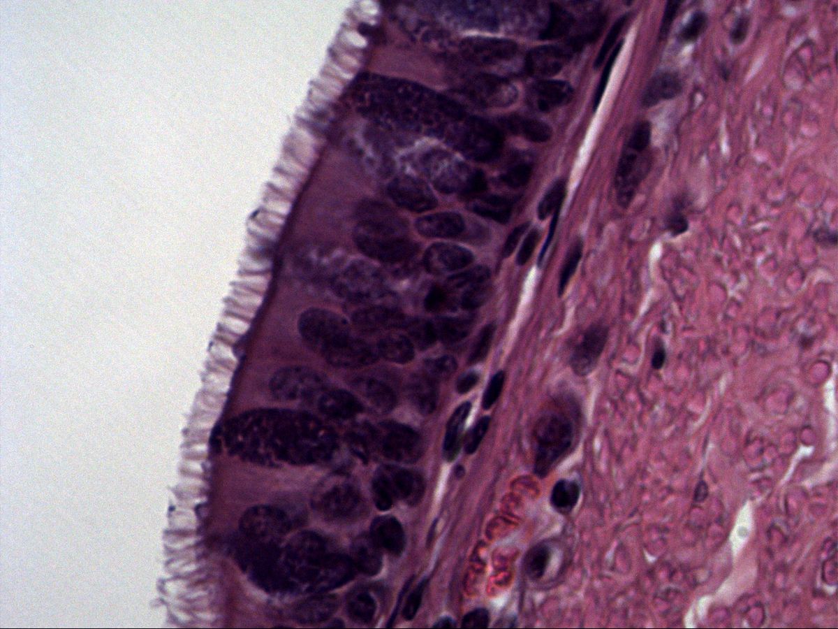
Pseudostratified columnar ciliated epithelium with kinocilia. Basal cells and cells positioned somewhat higher can be recognised by their nuclei. The ciliated epithelium carries kinocilia. In smokers the epithelium can transform into a squamous epithelium (metaplasia).
The respiratory epithelium is pseudostratified: all cells touch the basement membrane, but not all reach the lumen. The nuclei therefore lie at several levels and mimic a stratified epithelium. Typical features are the dense covering of kinocilia, the dark line of basal bodies beneath them and a strikingly thick basement membrane.
How to recognise it
- Nuclei at several levels, but all cells on the basement membrane
- Dense covering of kinocilia
- Dark line of basal bodies beneath the surface
- Thick, eosinophilic basement membrane
- Goblet cells between the ciliated cells
Structures to find
- Kinocilia
- Basal bodies
- Nuclei (different shapes and levels)
- Basal cells
- Goblet cells
- Basement membrane
- Lumen
Structure
The epithelium contains several cell types: columnar ciliated cells with kinocilia (about 5–7 µm long, microtubules in a 9×2+2 pattern), mucus-producing goblet cells, small basal cells acting as stem cells near the basement membrane, and occasional brush and neuroendocrine cells. The cilia are rooted in basal bodies (kinetosomes), which appear in H&E as a dark line just beneath the cell surface. Below the epithelium lies a thick, eosinophilic basement membrane, followed by the lamina propria with capillaries, elastic fibres and seromucous glands.
Function
Goblet cells and glands produce a mucus film in which inhaled particles and microbes are trapped. The cilia beat in a coordinated way towards the pharynx and move the mucus upwards at about 1 cm per minute – mucociliary clearance. Basal cells replace worn-out ciliated and goblet cells. Pseudostratified ciliated epithelium lines the nasal cavity, paranasal sinuses, larynx (except the vocal folds), trachea and bronchi. In the smaller bronchioles it becomes lower and simple, and goblet cells are replaced by club cells.
Microscopy tips
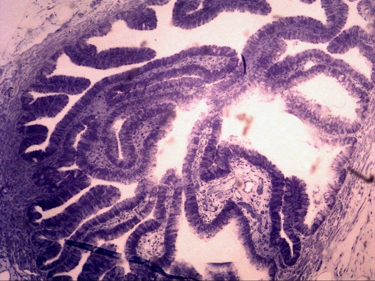
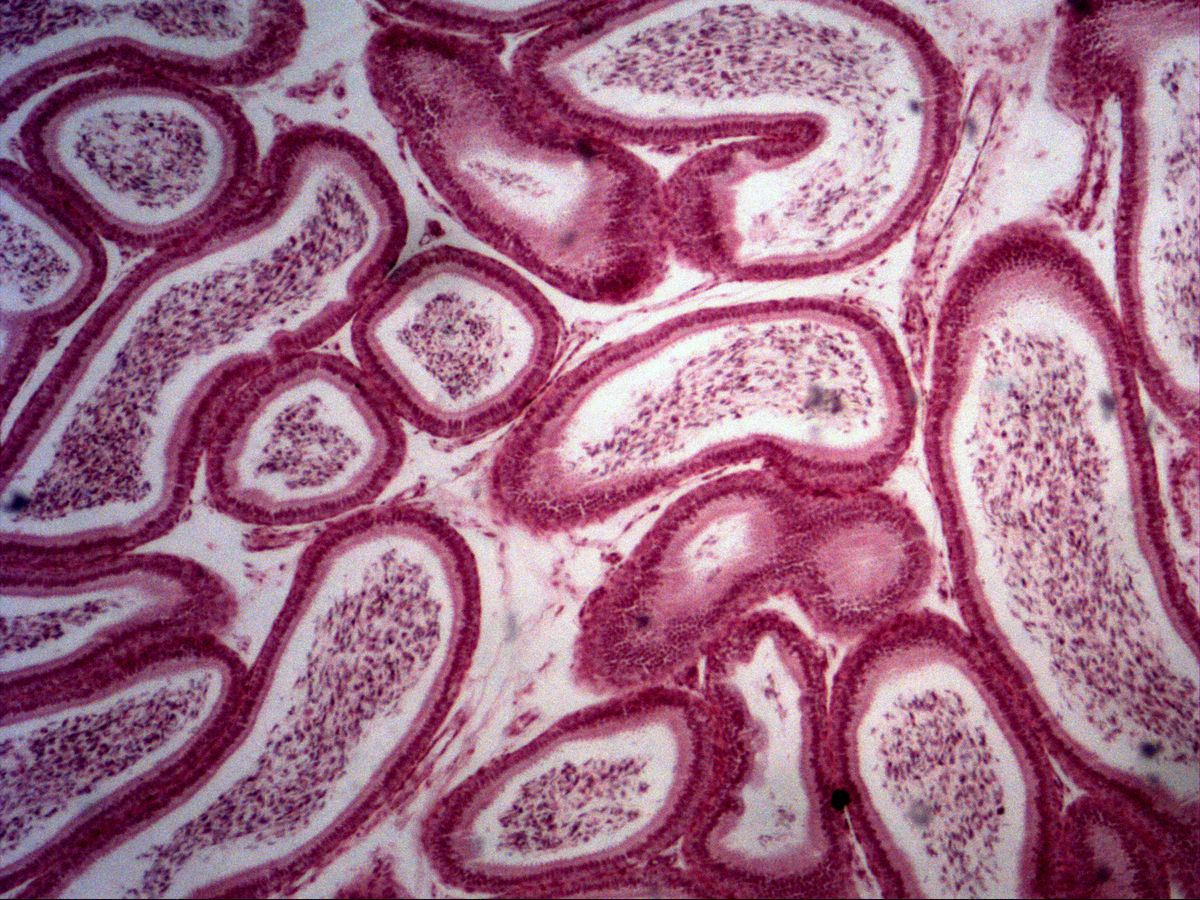
Find the mucosal surface at medium magnification, then switch to high power. Look for three things: the fine 'lawn' of cilia, the dark line of basal bodies and nuclei at different heights – the round nuclei of basal cells sit directly above the basement membrane, the oval nuclei of ciliated cells lie further apically. In H&E goblet cells appear as pale, goblet-shaped cells without cilia. Differential diagnosis: the epididymis carries non-motile, longer stereocilia without a basal body line; the uterine tube epithelium is simple.
Clinical relevance
Cigarette smoke paralyses the cilia and damages the epithelium. As an adaptation the ciliated epithelium can transform into stratified squamous epithelium (squamous metaplasia) – more resistant but without cleaning function. Bronchial carcinoma can develop from it. In primary ciliary dyskinesia (e.g. Kartagener syndrome) dynein arms are missing, leading to chronic respiratory infections. Because sperm flagella are affected as well, affected men are often infertile.
Test your knowledge
Assign the correct number in the image to each term.
Tap the requested structure in the image.
Multiple-choice questions on structure and function.
Frequently asked questions
What is the difference between pseudostratified and stratified?
In pseudostratified epithelium all cells reach the basement membrane; only the nuclei lie at different levels. In stratified epithelium only the lowest cell layer touches the basement membrane.
How do I tell cilia from microvilli?
Cilia are clearly longer, individually visible and sit on a dark line of basal bodies. Microvilli form only a short, homogeneous fringe without basal bodies.
Why is the tracheal basement membrane so thick?
Beneath the actual basal lamina lies a dense layer of reticular and collagen fibres (lamina fibroreticularis). Together they are visible in the light microscope as a broad pink band.
Related topics
Updated: