Goblet cells
Duodenum · trichrome
Labelled structures
This image has no labels – use the caption instead.
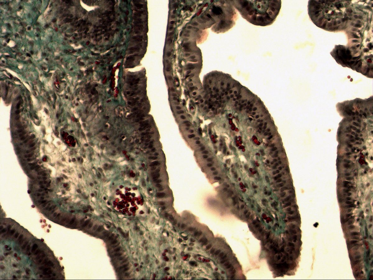
The goblet cells, widened apically into a cup shape, are easy to recognise. They are an intraepithelial, unicellular glandular tissue. Paneth cells, which are responsible for local immune defence, are also unicellular glands.
Goblet cells are the simplest example of an exocrine gland: a single epithelial cell produces mucins and releases them onto the surface by exocytosis. In the duodenum they are scattered between the absorptive enterocytes; in addition, the duodenum has a special feature, the multicellular duodenal (Brunner) glands in the submucosa.
How to recognise it
- Pale, cup-shaped distended cells in the surface epithelium
- Flattened nucleus in the narrow basal stem
- Lie singly between columnar enterocytes
- Neighbouring enterocytes with an eosinophilic brush border
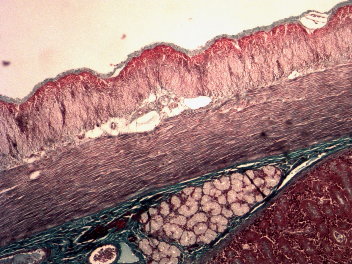
- Duodenum: mucous Brunner glands in the submucosa
Structures to find
- Goblet cells
- Enterocytes
- Brush border (microvilli)
- Different nuclei (goblet cell vs. enterocyte)
- Duodenal glands (Brunner glands)
- Submucosa
Structure
The goblet cell is a columnar epithelial cell with clear polarity: its narrow stem rests on the basement membrane and contains the flattened, often triangular nucleus, rough ER and Golgi apparatus. Apically, the cell is distended into a cup (theca) by densely packed mucin granules. In H&E or trichrome staining the granules remain largely unstained, so the cup looks pale and foamy. The neighbouring enterocytes are slender columnar cells with an elongated, basal nucleus and an eosinophilic brush border of microvilli.
Function
Goblet cells secrete mucins, heavily glycosylated proteins that swell with water to form a viscous mucus layer. This protects the epithelium mechanically and chemically, eases the passage of intestinal contents and keeps microorganisms at a distance. Secretion is merocrine; under strong stimulation the whole cup may be emptied at once. The number of goblet cells increases markedly from the duodenum towards the colon. The Brunner glands add to this protection: their alkaline, bicarbonate-rich mucus neutralises acidic gastric chyme.
Microscopy tips
At medium magnification look at the surface epithelium of the villi for pale, round to oval gaps between the darker enterocytes. At high magnification you can see that the cup opens towards the lumen and that the nucleus is compressed in the narrow stem below it. In the overview you recognise the duodenum by its Brunner glands: pale, mucous gland lobules beneath the muscularis mucosae. Pitfall: goblet cells cut in cross-section can resemble vacuoles or fat cells – what matters is their position within the epithelium.
Clinical relevance
In intestinal metaplasia of the gastric mucosa or in Barrett's oesophagus, goblet cells appear where they are normally absent – an important diagnostic criterion in pathology. In cystic fibrosis the mucus is thick because of defective chloride and water transport and can obstruct the bowel. Rarely, hyperplasia of the Brunner glands presents as a duodenal polyp. In inflammatory bowel disease such as ulcerative colitis, by contrast, goblet cell depletion is typical.
Slide images
Test your knowledge
Assign the correct number in the image to each term.
Tap the requested structure in the image.
Multiple-choice questions on structure and function.
Frequently asked questions
Why do goblet cells look pale in H&E?
The mucin granules mainly contain acidic glycoproteins that hardly take up haematoxylin or eosin. With PAS or Alcian blue staining, by contrast, they stain intensely.
Are goblet cells exocrine or endocrine?
Exocrine: they release their secretion directly onto the epithelial surface. They are classed as unicellular, intraepithelial exocrine glands.
How do I know the section is from the duodenum?
By the mucous Brunner glands in the submucosa. The jejunum and ileum have no such submucosal glands.
Related topics
Updated: