Learning path · Histology
Nervous tissue
Pseudounipolar, bipolar and multipolar neurons, Purkinje and pyramidal cells and the structure of peripheral nerves – with glia, Nissl substance and myelin sheaths.
-
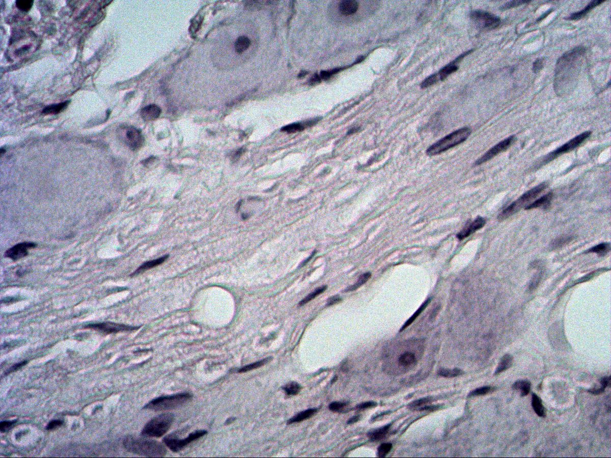
Pseudounipolar neurons
Trigeminal ganglion · H&E
4 images 14 structures -
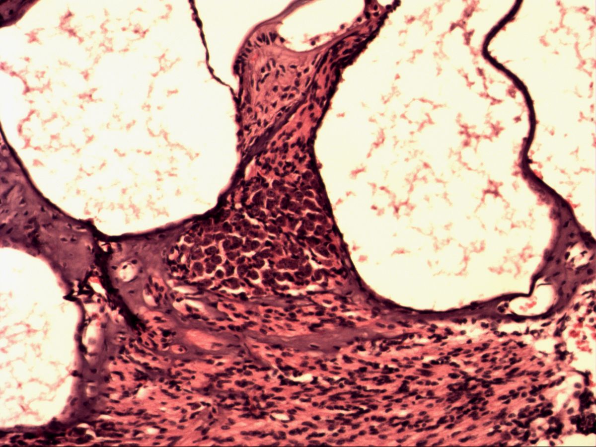
Bipolar neurons
Spiral ganglion (cochlea) · H&E
4 images 6 structures -
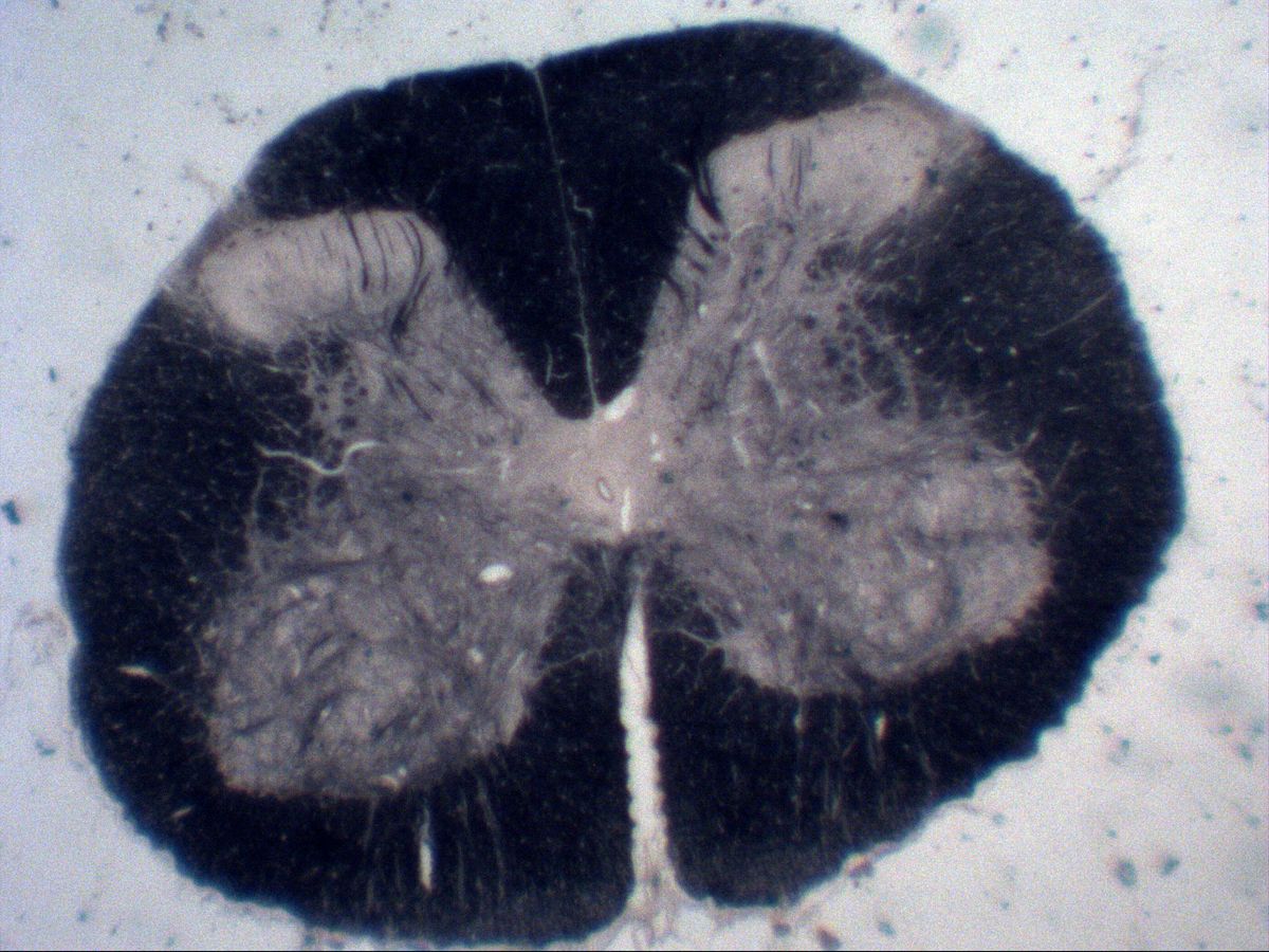
Multipolar neurons
Spinal cord · H&E / myelin stain
9 images 15 structures -
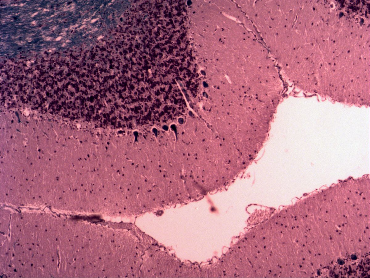
Purkinje cells
Cerebellum · Fast Blue / H&E
11 images 17 structures -
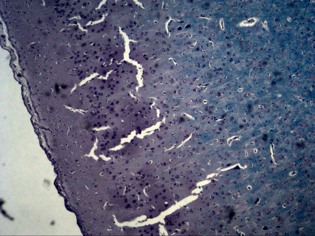
Pyramidal cells
Cerebral cortex · H&E / Fast Blue
7 images 19 structures -
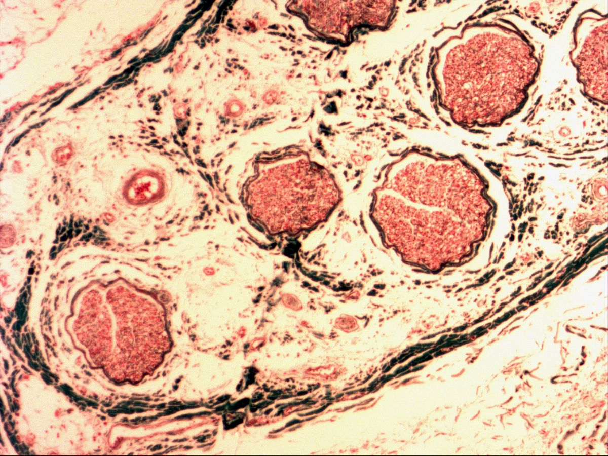
Peripheral nerve
Femoral nerve · trichrome
7 images 19 structures
Test your knowledge
Mixed exam simulation: identify slides, name highlighted structures and answer knowledge questions. Choose your areas and off you go.