Pyramidal cells
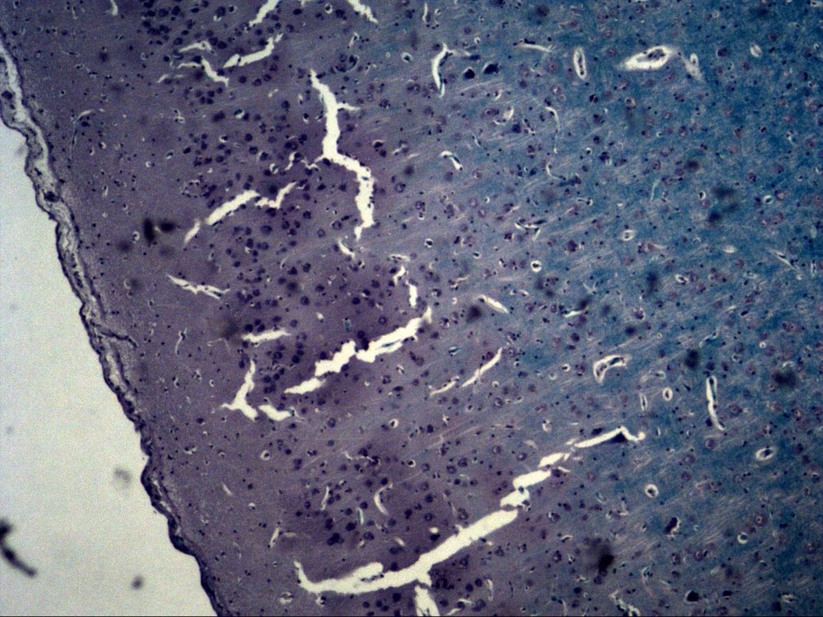
Cerebral cortex · H&E / Fast Blue
Labelled structures
This image has no labels – use the caption instead.
Pyramidal cells are the typical projection neurons of the cerebral cortex (isocortex). Their soma is triangular to cone-shaped: a long apical dendrite runs from the apex towards the brain surface, while several basal dendrites and the axon leave from the base. They are multipolar, glutamatergic and therefore excitatory neurons.
The images show cerebral cortex stained with H&E and Fast Blue: overviews with the brain surface and pia mater, individual pyramidal cells with nucleus and nucleolus, small glial nuclei in the neuropil and a capillary containing red blood cells.
How to recognise it
- Triangular to cone-shaped soma
- Thick apical dendrite at the apex, pointing towards the surface
- Large, pale nucleus with a prominent nucleolus
- Basophilic Nissl substance in the cytoplasm
- Fine fibrous neuropil with small, dark glial nuclei
- No dense granular layer and no single cell row as in the cerebellum
Structures to find
- Pyramidal cell (soma)
- Apical dendrite
- Nucleus with nucleolus
- Glial nuclei
- Neuropil
- Capillary with endothelial nucleus
- Pia mater at the brain surface
Structure of the cerebral cortex
The isocortex consists of six layers, numbered from the surface inwards:
- I Molecular layer: few cells, mainly fibres and dendrites
- II External granular layer: small granule and pyramidal cells
- III External pyramidal layer: medium-sized pyramidal cells
- IV Internal granular layer: granule cells, input from the thalamus
- V Internal pyramidal layer: large pyramidal cells, including the giant Betz cells of the motor cortex
- VI Multiform layer: cells of varied shape
The white matter lies beneath. The layer boundaries are indistinct and are hard to define reliably on H&E sections without practice.
Pyramidal cell and neuropil
The soma of a pyramidal cell contains a large, pale (euchromatic) nucleus with a prominent nucleolus and abundant basophilic Nissl substance (rough ER). The axons of pyramidal cells form the main projection pathways of the cortex: to other cortical areas, to the opposite hemisphere via the corpus callosum and to the brainstem and spinal cord (pyramidal tract). Between the neurons lies the neuropil, a fine meshwork of dendrites, axons and glial processes. In it you can see small, dark glial nuclei without visible cytoplasm, as well as capillaries. Their continuous endothelium with tight junctions, together with the basal lamina and astrocytic end-feet, forms the blood–brain barrier.
Microscopy tips
Start at low power and find the brain surface with the pia mater – this tells you which direction the apical dendrites should point. Then switch to high power: pyramidal cells are recognised by their triangular soma, a large pale nucleus with a nucleolus and a thick process at the apex. Glial nuclei are much smaller, round or elongated and darker. Clear spaces around neurons and vessels are usually shrinkage artefacts. Very dark, shrunken neurons (“dark neurons”) are often caused by tissue handling. Differential: cerebellar Purkinje cells lie in a row above a dense granular layer; spinal motor neurons are larger, polygonal and have coarse Nissl bodies.
Clinical relevance
Large pyramidal cells in layers III and V are particularly vulnerable to hypoxia. After a hypoxic insult they appear as “red neurons” with eosinophilic cytoplasm and a shrunken, dark nucleus. In amyotrophic lateral sclerosis the Betz cells of the motor cortex and the spinal motor neurons degenerate. In Alzheimer’s disease, pyramidal cells of the hippocampus and association cortex in particular lose their synapses and die. Damage to the pyramidal tract causes upper motor neuron paralysis with brisk reflexes and a positive Babinski sign.
Slide images
Test your knowledge
Assign the correct number in the image to each term.
Tap the requested structure in the image.
Multiple-choice questions on structure and function.
Frequently asked questions
Which layers contain most pyramidal cells?
Mainly in layer III (external pyramidal layer) and layer V (internal pyramidal layer). Layer V of the motor cortex contains the particularly large Betz cells, whose axons contribute to the pyramidal tract.
How do I distinguish neuronal from glial nuclei?
Neuronal nuclei are large, pale and round with a prominent nucleolus and are surrounded by visible cytoplasm. Glial nuclei are small, dark, often elongated or irregular and appear “naked” in the neuropil.
Are the clear halos around neurons and vessels real structures?
No, they are usually shrinkage artefacts caused by fixation and embedding. In the living brain, neuropil, neurons and vessels are closely apposed.
Related topics
Updated: