Bipolar neurons
Spiral ganglion (cochlea) · H&E
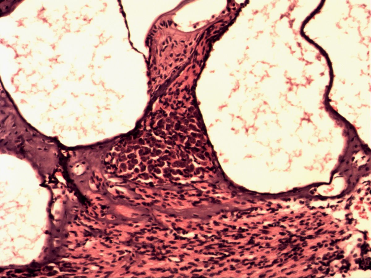
Labelled structures
This image has no labels – use the caption instead.
The spiral (cochlear) ganglion lies in the bony Rosenthal's canal at the base of the osseous spiral lamina and winds around the modiolus with the cochlea. It contains the perikarya of the bipolar neurons of the auditory nerve, which send one process to the hair cells of the organ of Corti and one to the brainstem.
How to recognise it
- Located in a bony canal at the base of the osseous spiral lamina
- Densely packed, oval perikarya
- Round pale nucleus with nucleolus
- Two processes at opposite poles
- Surrounding satellite or Schwann cells
Structures to find
- Spiral ganglion in the modiolus
- Perikaryon of a bipolar neuron
- Nucleus with nucleolus
- Dendritic and axonal pole (if sectioned)
- Schwann or satellite cells
- Osseous spiral lamina
- Cochlear nerve fibres
Structure
Bipolar neurons have an oval to spindle-shaped cell body with two processes at opposite poles: a dendritic (peripheral) pole and an axonal (central) pole. The nucleus is round, pale and euchromatic with a distinct nucleolus. The perikarya are smaller than those of pseudounipolar neurons, closely packed and surrounded by satellite cells; in many species (and largely in humans) Schwann cells even wrap the perikaryon in loose myelin. Nerve fibres run between the cell groups.
Function and occurrence
The bipolar neurons of the spiral ganglion are the first neuron of the auditory pathway. Their peripheral process runs through the osseous spiral lamina to the hair cells – about 95 % to the inner hair cells – and their central process forms the cochlear nerve, ending in the cochlear nuclei of the brainstem. Bipolar neurons also occur in the vestibular ganglion, in the retina (between photoreceptors and ganglion cells) and as olfactory receptor cells in the olfactory epithelium.
Microscopy tips
First orient yourself on the cochlea: the bony modiolus forms the axis, from which the osseous spiral lamina projects into the cochlear canal. At its base, in a bony canal, lies a dense nest of round cells – the spiral ganglion. At high magnification you see round, pale nuclei with a nucleolus in eosinophilic cytoplasm. Both processes are rarely caught in the same section; the label ‘bipolar’ follows mainly from the location. The perilymphatic spaces often contain precipitated, pink, stringy protein.
Clinical relevance
A cochlear implant bypasses lost hair cells and electrically stimulates the fibres and neurons of the spiral ganglion directly – it therefore only works if these neurons are preserved. A vestibular schwannoma (acoustic neuroma) in the internal acoustic meatus can compress the cochlear nerve, causing unilateral hearing loss. With age (presbycusis) and after noise damage, hair cells and, secondarily, spiral ganglion neurons are lost.
Slide images
Test your knowledge
Assign the correct number in the image to each term.
Tap the requested structure in the image.
Multiple-choice questions on structure and function.
Frequently asked questions
Where exactly is the spiral ganglion?
In Rosenthal's canal (spiral canal of the modiolus), a spiral bony canal in the modiolus at the base of the osseous spiral lamina.
How do bipolar neurons differ from pseudounipolar ones?
In bipolar neurons two separate processes leave opposite poles. In pseudounipolar neurons they fuse into one stem that divides in a T-shape.
Where else are bipolar neurons found?
In the vestibular ganglion, the retina and the olfactory epithelium.
Related topics
Updated: