Multipolar neurons
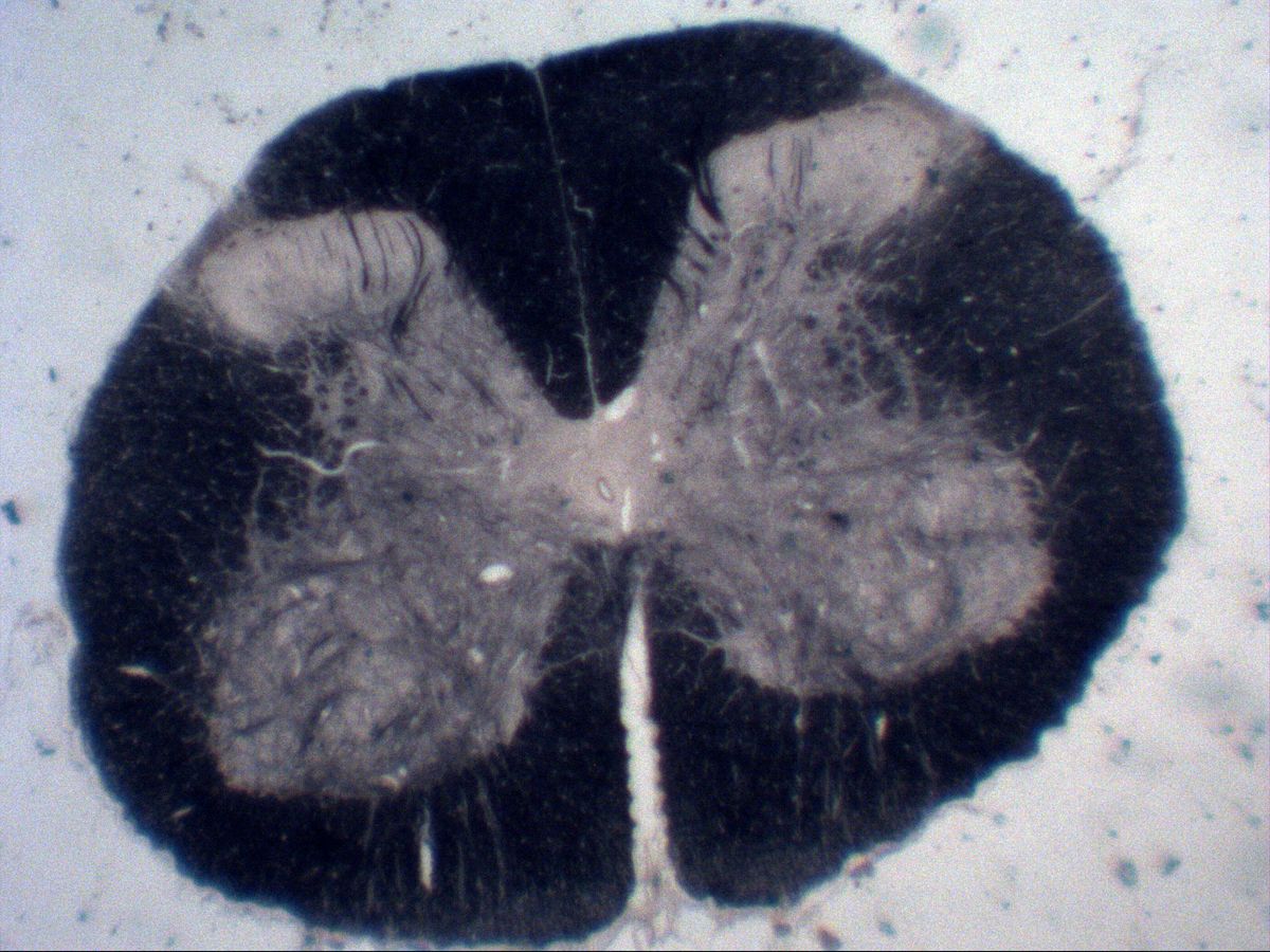
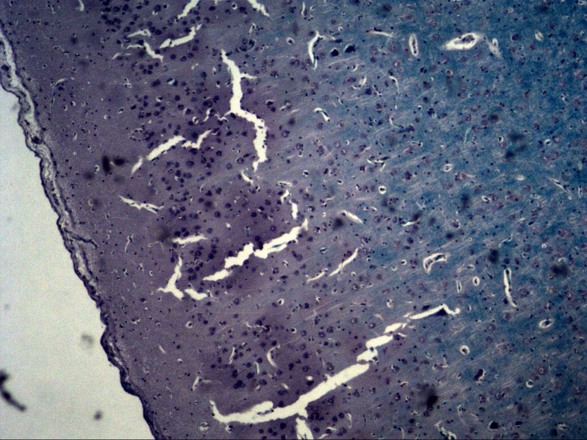
Spinal cord · H&E / myelin stain
Labelled structures
This image has no labels – use the caption instead.
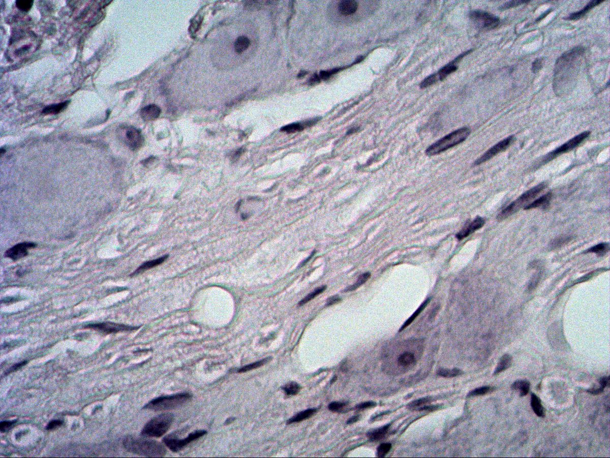
The ventral horn of the spinal cord contains the largest multipolar neurons of the body, the α-motor neurons. Their perikaryon gives rise to several dendrites and one axon. Different stains selectively show perikarya (H&E, Nissl), processes (silver impregnation) and myelin sheaths (myelin stain).
How to recognise it
- Large, polygonal perikarya in the ventral horn
- Several dendrites, one axon
- Coarse Nissl bodies, also in the dendrites
- Pale nucleus with distinct nucleolus
- Surrounding neuropil with astrocyte and oligodendrocyte nuclei
- Butterfly-shaped grey matter
Structures to find
- Grey matter with ventral and dorsal horn
- White matter
- Central canal with ependyma
- Anterior median fissure
- Perikaryon of a motor neuron
- Dendrites
- Axon hillock
- Nuclei of astrocytes and oligodendrocytes
Structure of the spinal cord
In cross-section the grey matter forms a butterfly-shaped figure around the central canal, which is lined by ependyma. The narrow dorsal horns receive sensory afferents; the broad ventral horns contain the motor neurons. Outside lies the white matter with ascending and descending myelinated tracts, divided into dorsal, lateral and ventral funiculi. Ventrally the wide anterior median fissure cuts in, dorsally the narrow posterior median septum. In myelin stains the white matter appears black and the grey matter pale.
Multipolar neurons
α-motor neurons have a large, polygonal perikaryon with a central pale nucleus and a distinct nucleolus. The cytoplasm is full of coarse basophilic Nissl bodies, which extend into the proximal dendrites. The axon hillock, where the axon originates, is free of Nissl substance and therefore appears pale. Around the neurons lies the neuropil of processes and glial cells: astrocytes with larger, pale, oval nuclei and oligodendrocytes with small, round, dark nuclei, often with a clear perinuclear halo.
Microscopy tips
In the overview, orient yourself by the shape of the grey matter: the ventral horns are broad and plump, the dorsal horns narrow and reach almost to the surface; the wide fissure lies ventrally. Then look for the motor neurons in the ventral horn at higher magnification. A dendrite is a tapering process containing Nissl bodies; the axon arises from the pale axon hillock – it is rarely sectioned. In silver impregnation neurofibrils and processes appear black; Nissl stains (e.g. cresyl violet) show only perikarya and nuclei. Clear spaces around perikarya are shrinkage artefacts.
Clinical relevance
In amyotrophic lateral sclerosis (ALS) the ventral horn motor neurons and the corticospinal tract degenerate; in poliomyelitis the ventral horn cells are destroyed by the virus. If an axon is severed, the perikaryon reacts with chromatolysis: it swells, the Nissl bodies dissolve and the nucleus moves to the periphery. Spinal cord injuries interrupt the white matter tracts and cause paralysis and sensory loss below the lesion.
Slide images
Test your knowledge
Assign the correct number in the image to each term.
Tap the requested structure in the image.
Multiple-choice questions on structure and function.
Frequently asked questions
How do I tell a dendrite from the axon in a section?
Dendrites contain Nissl bodies and taper gradually. The axon arises from the pale, Nissl-free axon hillock and has a uniform diameter.
Why is the white matter black in a myelin stain?
The stain binds to myelin lipids. White matter consists mainly of myelinated fibres, grey matter mainly of perikarya and neuropil.
How do I recognise oligodendrocytes and astrocytes in H&E?
Oligodendrocytes have small, round, dark nuclei, often with a clear halo. Astrocyte nuclei are larger, more oval and paler with fine chromatin; their cytoplasm is barely visible in H&E.
Related topics
Updated: