Pseudounipolar neurons
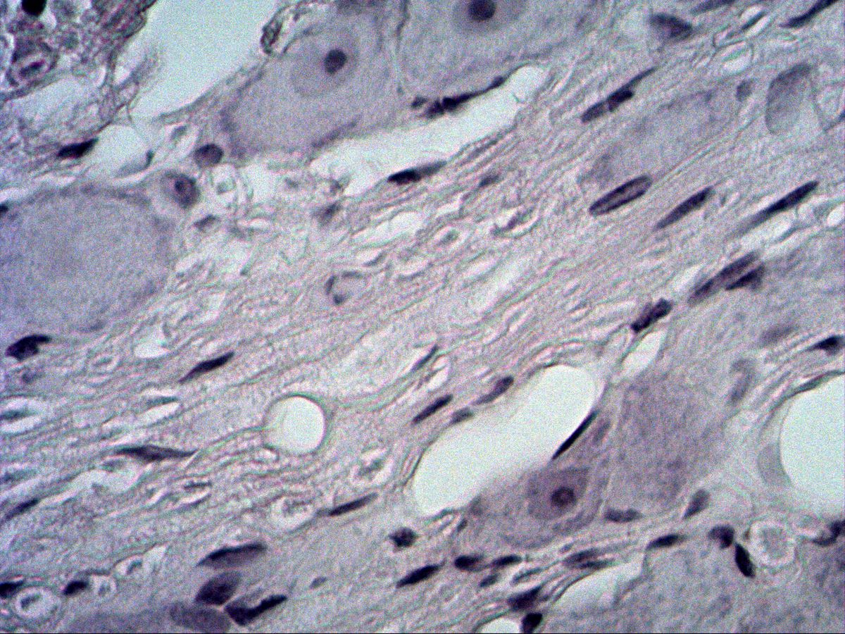
Trigeminal ganglion · H&E
Labelled structures
This image has no labels – use the caption instead.
The trigeminal ganglion is a sensory ganglion containing the perikarya of the pseudounipolar neurons of the trigeminal nerve. Sections show large, round nerve cell bodies, each surrounded by a ring of small satellite (mantle) cells, with bundles of nerve fibres in between.
How to recognise it
- Large, round perikarya
- Pale euchromatic nucleus with a large nucleolus
- Fine Nissl bodies in the cytoplasm
- Ring of small satellite cell nuclei around each perikaryon
- Nerve fibre bundles between the cell groups
Structures to find
- Perikaryon of a pseudounipolar neuron
- Nucleus
- Nucleolus
- Nissl bodies
- Satellite (mantle) cells
- Axon hillock or initial process (if sectioned)
- Nerve fibres with Schwann cell nuclei
Structure
Pseudounipolar neurons have a large, round perikaryon (up to about 100 µm) with a large, pale, euchromatic nucleus and a prominent dark nucleolus (‘owl's eye’ appearance). The cytoplasm contains fine Nissl bodies (rough ER) and, in older individuals, brownish lipofuscin. A single process leaves the cell body and divides in a T-shape into a peripheral and a central branch. Each perikaryon is enveloped by flat satellite (mantle) cells (peripheral glia) and a basal lamina, followed by a thin connective tissue layer.
Function and occurrence
Pseudounipolar neurons are primary afferent, sensory neurons. Their peripheral branch conducts information from receptors in skin, mucosa, teeth and muscles to the ganglion; the central branch carries it on into the CNS – for the trigeminal nerve into the brainstem. The impulse bypasses the perikaryon, which mainly sustains the long processes. Occurrence: spinal ganglia and sensory cranial nerve ganglia (V, VII, IX, X).
Microscopy tips
At low magnification you can see groups of large, round cells between tracts of nerve fibres. At high magnification look for the pale nucleus with a large nucleolus and the ring of small dark satellite cell nuclei immediately around the perikaryon. The nucleus is only visible if the cell is cut through its centre – many perikarya therefore appear anucleate. The initial process or axon hillock is rarely caught in the section. Versus an autonomic ganglion: there the neurons are multipolar, smaller, often have an eccentric nucleus and fewer satellite cells.
Clinical relevance
Varicella zoster virus persists in sensory ganglia after chickenpox. If it reactivates in the trigeminal ganglion, herpes zoster ophthalmicus results, with a painful vesicular rash in the territory of the ophthalmic nerve. Herpes simplex viruses (cold sores) also remain latent here. In trigeminal neuralgia, sudden, severe stabbing facial pain occurs, often caused by a vessel compressing the nerve root. For treatment, the trigeminal ganglion can be reached through the foramen ovale with a needle and treated with heat or balloon compression.
Slide images
Test your knowledge
Assign the correct number in the image to each term.
Tap the requested structure in the image.
Multiple-choice questions on structure and function.
Frequently asked questions
Why are the cells called ‘pseudounipolar’?
They develop from bipolar precursors whose two processes fuse into a common stem. This divides in a T-shape into a peripheral and a central branch.
What are satellite (mantle) cells?
Flat peripheral glial cells that completely envelop each perikaryon in the ganglion. They support and nourish the neuron and regulate its microenvironment.
Why do many neurons show no nucleus?
The perikarya are much larger than the section thickness. Only sections through the centre of the cell include the nucleus.
Related topics
Updated: