Purkinje cells
Cerebellum · Fast Blue / H&E
Labelled structures
This image has no labels – use the caption instead.
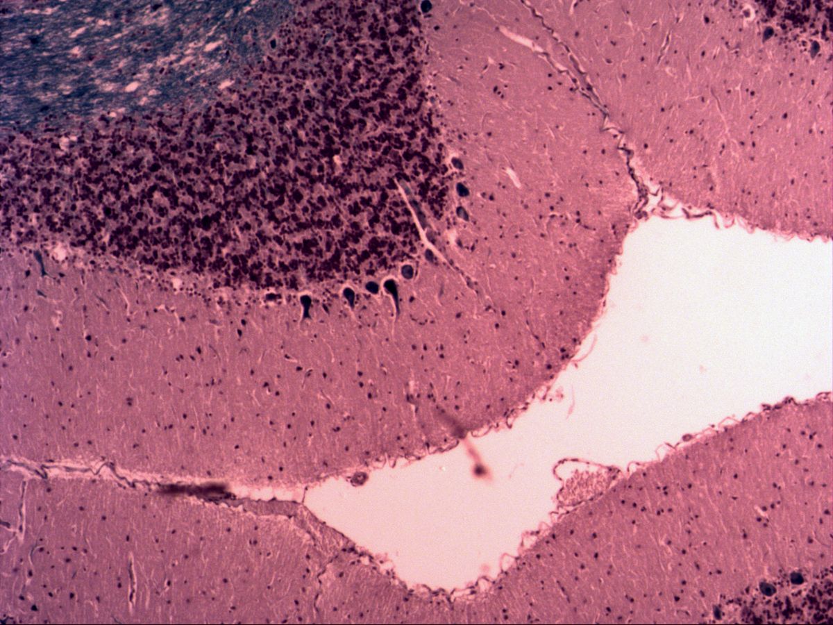
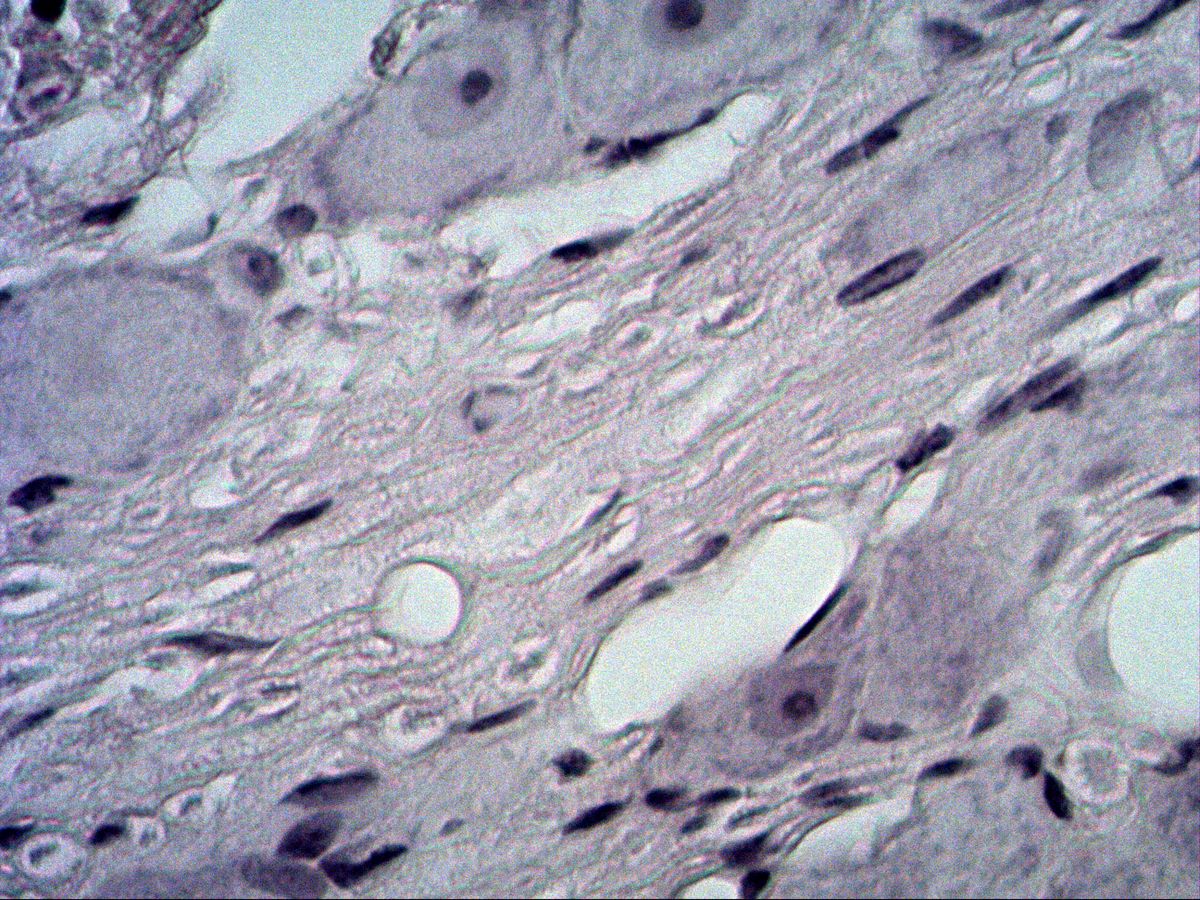
Purkinje cells are the largest neurons of the cerebellar cortex and its only efferent neurons. Their pear-shaped cell bodies form a single row at the boundary between the cell-poor molecular layer and the cell-rich granular layer. A huge dendritic tree branches like an espalier in one plane, perpendicular to the long axis of the folium, almost up to the surface.
The images show cerebellar cortex stained with Fast Blue (myelin blue, nuclei and Nissl substance violet) and with an H&E or Klüver-Barrera-type stain. The three-layered cortex is visible even at overview magnification; at high magnification you can see the soma, the nucleus with its nucleolus and the main dendritic trunk.
How to recognise it
- Three-layered cortex: pale molecular layer, dark granular layer, a row of cells in between
- Large, pear-shaped cell bodies in a single row (Purkinje cell layer)
- Thick dendritic trunk running into the molecular layer towards the surface
- Large, pale nucleus with a prominent nucleolus
- Granular layer of densely packed, small, round nuclei
- Leaf-like folds (folia) with central white matter
Structures to find
- Molecular layer
- Purkinje cell layer (ganglionic layer)
- Granular layer
- White matter
- Soma (perikaryon) of a Purkinje cell
- Nucleus with nucleolus
- Dendritic tree with main trunk
Structure
The cerebellar cortex (cortex cerebelli) has the same three layers everywhere:
- Molecular layer (outer): few cells, many fibres; contains the dendritic trees of Purkinje cells, the parallel fibres of granule cells and stellate and basket cells.
- Purkinje cell layer (ganglionic layer): a single row of large Purkinje cell bodies with Bergmann glial cells in between.
- Granular layer (inner): densely packed, small, round nuclei of granule cells, plus Golgi cells and nucleus-free islands (cerebellar glomeruli).
Beneath lies the white matter with myelinated afferents (mossy and climbing fibres) and the axons of the Purkinje cells.
Function
Purkinje cells are GABAergic, i.e. inhibitory, projection neurons. The axon leaves the basal pole of the soma, passes through the granular layer and white matter and ends on the deep cerebellar nuclei (e.g. the dentate nucleus) and the vestibular nuclei. Each Purkinje cell receives two very different excitatory inputs: countless parallel fibres from granule cells, which are themselves driven by mossy fibres, and a single climbing fibre from the inferior olive that winds up along the dendritic tree. By comparing these signals the cerebellum corrects ongoing movements. It is therefore essential for coordination, balance, muscle tone and motor learning.
Microscopy tips
At low power the cerebellum is recognised by its leaf-like folds (folia): white matter in the centre, a dark band of nuclei around it (granular layer) and a broad, pale outer zone (molecular layer). Then follow the boundary between the granular and molecular layers – this is where the Purkinje cell bodies sit, often spaced apart rather than in a continuous line. At high power look for the large, pale nucleus with a prominent nucleolus and the thick dendritic trunk heading towards the surface. With Fast Blue the white matter is blue. Pitfall: the cerebral cortex has no dense granular band and no single row of cells at a sharp layer boundary; its pyramidal cells are spread over several layers.
Clinical relevance
Purkinje cells are highly sensitive to hypoxia and toxic damage. Chronic alcohol misuse typically causes atrophy of the cerebellar vermis with loss of Purkinje cells and a broad-based ataxic gait. Purkinje cells are also lost in hereditary spinocerebellar ataxias and in paraneoplastic syndromes (e.g. with anti-Yo antibodies). Histologically there are gaps in the Purkinje cell row and an increase in Bergmann glia. Clinically, cerebellar damage presents with ataxia, intention tremor, dysmetria, dysdiadochokinesia and scanning speech.
Slide images
Test your knowledge
Assign the correct number in the image to each term.
Tap the requested structure in the image.
Multiple-choice questions on structure and function.
Frequently asked questions
Why is the inner layer called the granular layer?
It consists of vast numbers of tiny granule cells whose small, round, dark nuclei are packed so densely that they look like grains at low power. Granule cells are the most numerous neurons in the entire brain.
Where does the axon of a Purkinje cell go?
It arises from the basal pole of the soma, passes through the granular layer and white matter and ends with inhibitory synapses on the deep cerebellar nuclei or vestibular nuclei. Purkinje cells are therefore the sole output of the cerebellar cortex.
How do I tell cerebellar from cerebral cortex on a slide?
The cerebellum shows folia with a dense, dark granular band and a single row of large Purkinje cells along its outer edge. The cerebral cortex looks more uniform, has six poorly demarcated layers and triangular pyramidal cells at various depths.
Related topics
Updated: