Peripheral nerve
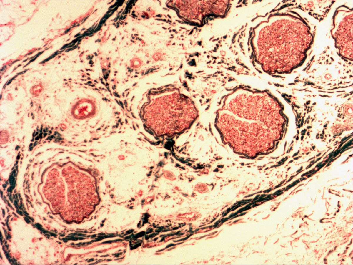
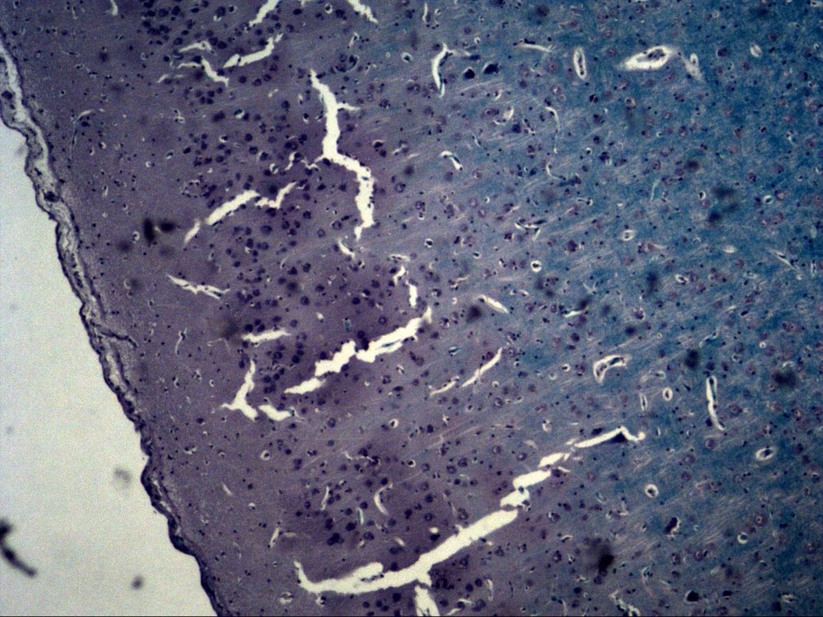
Femoral nerve · trichrome
Labelled structures
This image has no labels – use the caption instead.
A peripheral nerve is a bundle of many nerve fibres organised by three connective tissue sheaths: the epineurium surrounds the whole nerve, the perineurium each fascicle and the endoneurium each individual nerve fibre. Most fibres of a mixed nerve such as the femoral nerve are myelinated: the axon is wrapped in a myelin sheath formed by Schwann cells.
The images show the femoral nerve stained with trichrome (collagen green, axons and cytoplasm red) in cross-section with several fascicles, at high magnification with individual myelinated fibres, and in longitudinal section.
How to recognise it
- Round fascicles sharply bounded by the thin perineurium
- Loose, vascular epineurium between the fascicles
- Small circles in cross-section: red axon in the centre, clear myelin ring around it
- Fine green collagen fibres (endoneurium) between the fibres
- Fibres of different diameters side by side
- Wavy fibre course with elongated Schwann cell nuclei in longitudinal section
Structures to find
- Epineurium
- Perineurium
- Endoneurium
- Fascicle (nerve fibre bundle)
- Blood vessels (vasa nervorum)
- Axon
- Myelin sheath (appears white)
- Schwann cell nucleus
- Thick and thin axons
Structure
- Epineurium: loose to dense connective tissue with adipocytes and larger blood vessels (vasa nervorum); holds the fascicles together and forms a firmer outer layer around the nerve.
- Perineurium: several layers of flattened perineurial cells joined by tight junctions and covered by a basal lamina; surrounds each fascicle as a thin, sharply defined sheath.
- Endoneurium: delicate loose connective tissue with capillaries between the individual nerve fibres.
- Nerve fibre: axon plus a sheath of Schwann cells. In myelinated fibres each Schwann cell forms one internode of the myelin sheath, with a node of Ranvier between two internodes. Unmyelinated fibres lie in groups embedded in a single Schwann cell.
Function
The myelin sheath insulates the axon electrically. Action potentials are therefore generated only at the nodes of Ranvier and “jump” from node to node (saltatory conduction). The thicker the axon and myelin sheath, the faster the conduction: thick fibres (e.g. for motor control and proprioception) conduct at up to about 100 m/s, thin unmyelinated fibres (e.g. for pain and autonomic signals) at only around 1 m/s. The perineurium forms a diffusion barrier (blood–nerve barrier) that protects the internal milieu of the fascicles. Epineurium and perineurium make the nerve resistant to tension yet flexible.
Microscopy tips
At low power look for round fascicles sharply outlined by a thin, dark line (perineurium) and embedded in green, loose epineurium with vessels. At high power the cross-section shows many small circles: the red axon lies in the centre, surrounded by a clear ring – the myelin is largely dissolved during paraffin embedding and therefore looks white. Fine green collagen fibres in between belong to the endoneurium. In longitudinal section the fibres run in a slightly wavy course and the Schwann cell nuclei are elongated. Pitfall: tendon and smooth muscle lack clear myelin rings around a central axon and have no fascicles bounded by perineurium.
Clinical relevance
When an axon is transected, its distal part degenerates (Wallerian degeneration). If the endoneurial tubes remain intact, Schwann cells form guiding bands along which the axon regrows at about 1–3 mm per day. In Guillain–Barré syndrome an autoimmune reaction attacks the myelin sheaths and slows conduction. Diabetic polyneuropathy mainly damages long fibres and presents with stocking-type sensory loss. A femoral nerve lesion weakens the quadriceps femoris (knee extension) and abolishes the patellar reflex.
Slide images
Test your knowledge
Assign the correct number in the image to each term.
Tap the requested structure in the image.
Multiple-choice questions on structure and function.
Frequently asked questions
Why does the myelin sheath look white on the slide?
Myelin consists mainly of lipids, which are dissolved during dehydration and paraffin embedding. What remains is a clear ring around the axon, often containing only a fine protein meshwork (neurokeratin).
What is the difference between perineurium and endoneurium?
The perineurium wraps an entire fascicle and consists of flattened, epithelium-like joined cells that form a diffusion barrier. The endoneurium is delicate connective tissue surrounding each individual nerve fibre within the fascicle.
How do I recognise Schwann cell nuclei?
In cross-section they appear as flat to crescent-shaped nuclei hugging the outside of the myelin ring; in longitudinal section they are elongated and aligned with the fibre. With the light microscope they often cannot be reliably distinguished from endoneurial fibroblast nuclei.
Related topics
Updated: