Retina
Eye
Labelled structures
This image has no labels – use the caption instead.
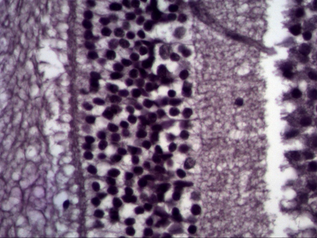
The retina is an outpost of the brain. Its light-sensitive part (pars optica) is strictly layered: three nuclear layers containing the cell bodies of photoreceptors, interneurons and ganglion cells alternate with two plexiform (synaptic) layers. Light has to pass through all the inner layers before reaching the photoreceptors on the outside (inverted retina).
The image is an older capture at high magnification. It shows a broad nuclear layer next to a finely reticular plexiform layer; exactly which layers are involved can only be stated with reservation without an overview image.
How to recognise it
- Strictly parallel layering across the whole width
- Three dark nuclear layers alternating with two pale, finely reticular plexiform layers
- Outer nuclear layer: densely packed, small, dark nuclei
- Inner plexiform layer clearly wider than the outer one
- Pigment epithelium and choroid outside, vitreous space inside
- Ganglion cell layer as a single row of larger cells
Structures to find
- Pigment epithelium
- Layer of rods and cones
- Outer nuclear layer
- Outer plexiform layer
- Inner nuclear layer
- Inner plexiform layer
- Ganglion cell layer
- Nerve fibre layer
Structure
From the outside (choroid) to the inside (vitreous body) ten layers are distinguished:
- Pigment epithelium
- Layer of rods and cones (outer and inner segments)
- External limiting membrane
- Outer nuclear layer: photoreceptor nuclei
- Outer plexiform layer: photoreceptor–bipolar cell synapses
- Inner nuclear layer: nuclei of bipolar, horizontal, amacrine and Müller cells
- Inner plexiform layer: bipolar cell–ganglion cell synapses
- Ganglion cell layer
- Nerve fibre layer (ganglion cell axons)
- Internal limiting membrane
Function
Signal processing follows a chain of three neurons: photoreceptor → bipolar cell → ganglion cell. Rods mediate night vision, cones colour and sharp vision. Horizontal cells (in the outer plexiform layer) and amacrine cells (in the inner plexiform layer) provide lateral connections and enhance contrast. Ganglion cell axons converge to form the optic nerve. Müller cells are glial cells that span the entire retina and form both limiting membranes. The pigment epithelium phagocytoses shed outer segment discs, regenerates visual pigment and absorbs stray light.
Microscopy tips
First orient yourself at low power: the brownish-black pigment epithelium and the vascular choroid lie on the outside, the vitreous space on the inside. Then count the three dark nuclear bands. The outer nuclear layer is usually the densest, with small, very dark, round nuclei. The inner nuclear layer is looser and contains nuclei of varying size. The ganglion cell layer consists of a single row of larger cells. The inner plexiform layer is clearly wider than the outer one. Common artefacts are a cleft between the pigment epithelium and the photoreceptors that mimics retinal detachment, and splits within the retina. Without an overview, a highly magnified field often cannot be assigned with certainty.
Clinical relevance
In retinal detachment the neural retina separates from the pigment epithelium along the former cleft between the two layers of the optic cup. Without contact with the pigment epithelium the photoreceptors die, so it is an emergency. Age-related macular degeneration damages the pigment epithelium and cones of the macula and impairs central vision. Diabetic retinopathy affects the capillaries of the inner retina, causing microaneurysms, haemorrhages and new vessel formation. In retinitis pigmentosa the rods are lost first – night blindness and constriction of the visual field are typical.
Test your knowledge
Assign the correct number in the image to each term.
Tap the requested structure in the image.
Multiple-choice questions on structure and function.
Frequently asked questions
Why is the retina called “inverted”?
Because the light-sensitive photoreceptors lie on the outermost side, facing away from the light. Light first has to pass through all the inner layers, which are largely transparent.
How do I tell the outer from the inner nuclear layer?
The outer nuclear layer lies closer to the pigment epithelium and consists of very densely packed, uniformly small, dark nuclei. The inner nuclear layer lies between the two plexiform layers and contains more loosely arranged nuclei of varying size.
Is a gap between the pigment epithelium and photoreceptors on a slide a retinal detachment?
Usually not: this boundary is mechanically weak and often tears during processing. It is then an artefact – though it lies exactly where a genuine detachment occurs.
Related topics
Updated: