Endocrine gland
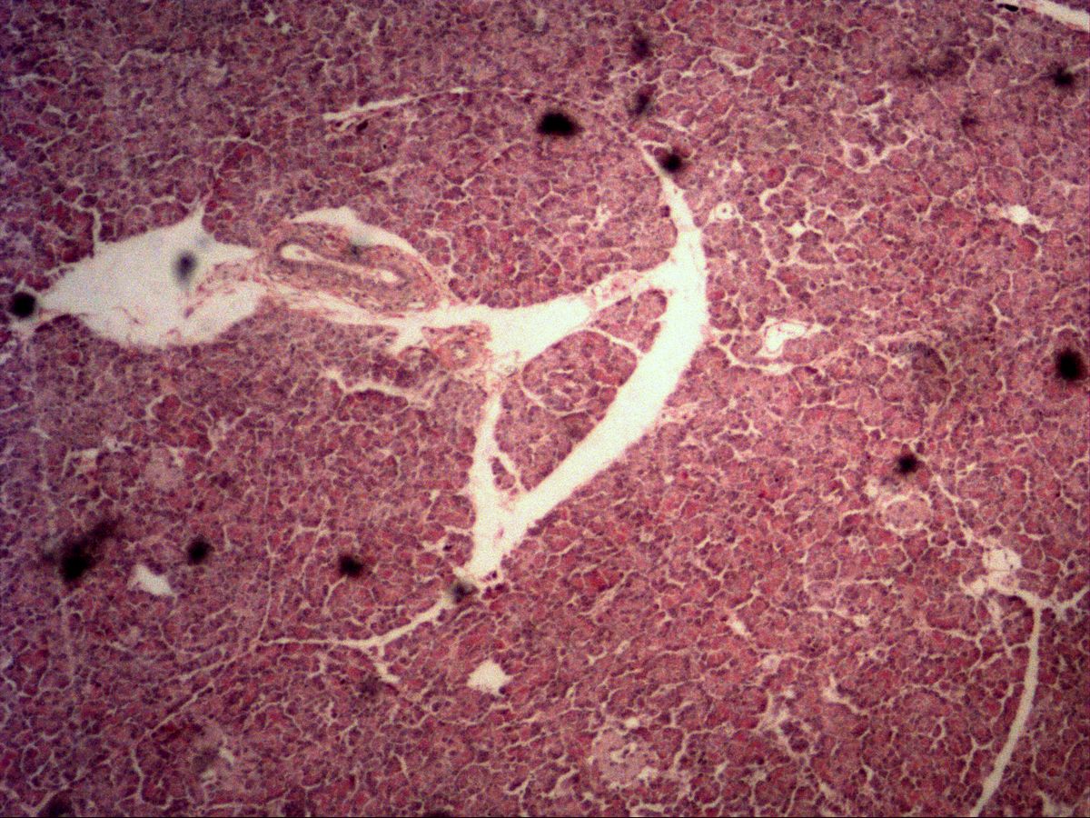
Pancreas · H&E
Labelled structures
This image has no labels – use the caption instead.
Endocrine glands release their secretion directly into the blood vessels. This is also the case for the islets of Langerhans: the endocrine glands of the pancreas mainly secrete insulin and glucagon for the antagonistic regulation of blood glucose.
The pancreas combines two glands in one organ: the exocrine part (purely serous acini) makes up about 98 %, the roughly one million islets of Langerhans only 1–2 %. The islets are spherical cell clusters without a lumen or duct, permeated by a dense network of fenestrated capillaries – the typical construction of an endocrine gland.
How to recognise it
- Pale, round cell islands in the darker exocrine tissue
- No lumen and no duct
- Cell cords with a dense capillary network
- Pale, evenly stained cytoplasm
- Round nuclei with pale euchromatin
Structures to find
- Islet of Langerhans
- Capillaries within the islet
- Exocrine serous acini
- Interlobular connective tissue
- Nucleus with pale euchromatin
Structure
An islet of Langerhans is a roundish nest of cords of epithelial cells surrounded by a delicate connective tissue capsule. Islet cells are smaller than acinar cells; their cytoplasm is pale eosinophilic and evenly stained, and their nuclei are round with mostly pale euchromatin. Numerous capillaries run between the cell cords. Immunohistochemistry distinguishes B cells (about 70 %, insulin, mostly central), A cells (about 20 %, glucagon, mostly peripheral), D cells (somatostatin) and PP cells (pancreatic polypeptide). In H&E staining these cell types cannot be reliably distinguished.
Function
Insulin lowers blood glucose by promoting glucose uptake into muscle and fat cells and the synthesis of glycogen and fat. Glucagon acts as its antagonist and raises blood glucose via glycogenolysis and gluconeogenesis in the liver. Somatostatin inhibits the release of both hormones in a paracrine manner. The hormones are released by exocytosis into the interstitium and pass through the fenestrated capillaries directly into the blood, reaching the liver first via the portal vein. In contrast, the exocrine acini secrete digestive enzymes into a duct system leading to the duodenum.
Microscopy tips
At low magnification, look within the dark purple exocrine tissue for pale, round islets of varying size. At higher magnification confirm the islet: no lumen, no acinar arrangement, evenly pale cytoplasm, many capillaries. The acinar cells, by contrast, show clear polarity with a basophilic base and eosinophilic zymogen granules at the apex. Further clues for the pancreas: centroacinar cells in the acinar lumen and the absence of striated ducts (distinguishing it from the parotid).
Clinical relevance
In type 1 diabetes mellitus an autoimmune reaction destroys the B cells, so insulin must be replaced for life. In type 2 diabetes insulin resistance is the main problem; later the B cells become exhausted and amyloid is deposited in the islets. Rare neuroendocrine tumours of the islets, such as insulinoma, cause recurrent hypoglycaemia through uncontrolled hormone release. After pancreatectomy both exocrine and endocrine function are lost, so enzymes and insulin must be replaced.
Slide images
Test your knowledge
Assign the correct number in the image to each term.
Tap the requested structure in the image.
Multiple-choice questions on structure and function.
Frequently asked questions
Why are the islets paler than the acini in H&E?
Islet cells lack the extensive basophilic rough ER and the strongly eosinophilic zymogen granules of acinar cells. Their cytoplasm therefore stains evenly pale.
Can A and B cells be distinguished in H&E?
No, this requires special stains or immunohistochemistry for insulin and glucagon. In H&E you can only recognise the islet as a whole.
What distinguishes an endocrine from an exocrine gland?
Endocrine glands have no duct and release their hormones into the blood via capillaries. Exocrine glands conduct their secretion through ducts onto a surface or into a hollow organ.
Related topics
Updated: