Simple tubular gland
Colon · H&E
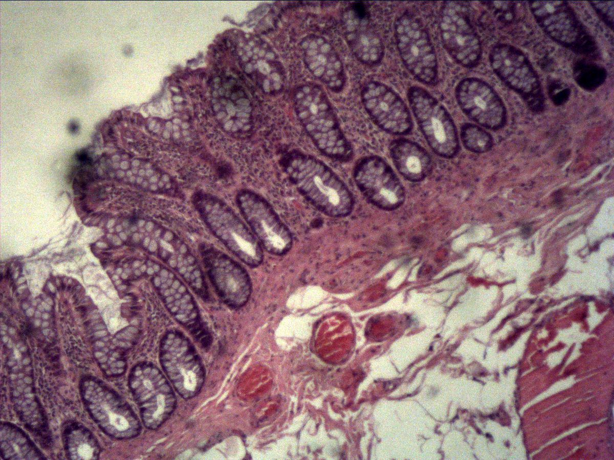
Labelled structures
This image has no labels – use the caption instead.
The test-tube-shaped glands also contain goblet cells.
The crypts of the large intestine (intestinal glands, crypts of Lieberkühn) are the classic example of a simple, unbranched tubular gland: a straight epithelial tube extends from the surface down to the muscularis mucosae. The colon has no villi, so the crypts stand close together like test tubes in a rack.
How to recognise it
- Straight, unbranched, test-tube-shaped crypts
- No villi, smooth mucosal surface
- Very many pale goblet cells in the crypt epithelium
- Narrow central lumen containing secretion
- Cell-rich lamina propria between the crypts
Structures to find
- Test-tube-shaped crypt (tubular gland)
- Goblet cells
- Nuclei of the crypt epithelium
- Secretion in the crypt lumen
- Basement membrane
- Lamina propria
Structure
Each crypt is a straight, blind-ending tube of simple columnar epithelium resting on a basement membrane. Most of its cells are goblet cells; between them lie absorptive cells (colonocytes), and at the crypt base stem cells and scattered enteroendocrine cells. In longitudinal section you see the test tube with a narrow central lumen; in cross-section, round profiles whose goblet cells form a rosette around the lumen. Between the crypts lies the cell-rich lamina propria with lymphocytes, plasma cells and capillaries.
Function
The main task of the colonic crypts is to produce mucus, which lubricates the increasingly dehydrated faeces and protects the mucosa from bacteria. The surface absorptive cells take up water and electrolytes. Stem cells divide at the crypt base; their daughter cells migrate towards the surface and are shed there after about five days. In this way the intestinal epithelium is constantly renewed. The secretion is released by merocrine secretion into the crypt lumen and from there into the gut lumen.
Microscopy tips
Start at low magnification: the colon shows a smooth surface without villi and straight crypts standing in parallel. At medium magnification the many pale goblet cells stand out and give the crypts a foamy appearance. To distinguish: the small intestine has villi and far fewer goblet cells, while the stomach has gastric pits with uniformly pale surface epithelium but no goblet cells. Obliquely cut crypts appear oval – trace them mentally down to the muscularis mucosae.
Clinical relevance
Most colorectal carcinomas arise from the crypts via adenomas, i.e. benign glandular neoplasms with dysplastic epithelium. In ulcerative colitis the crypts are branched and irregular, contain crypt abscesses and fewer goblet cells. These deviations from the normally regular test-tube shape are an important diagnostic criterion in biopsies. Polyps removed during screening colonoscopy are likewise assessed by whether their crypt epithelium still matures normally, i.e. forms goblet cells towards the surface, or whether dysplastic cells with enlarged, crowded nuclei reach the surface.
Slide images
Test your knowledge
Assign the correct number in the image to each term.
Tap the requested structure in the image.
Multiple-choice questions on structure and function.
Frequently asked questions
Why is it called a 'simple tubular' gland?
'Simple' means unbranched, and 'tubular' describes the tube-shaped secretory portion. There is no duct; the crypt opens directly onto the surface.
How do I tell colon from small intestine in a section?
The colon has no villi and very many goblet cells in long, straight crypts. The small intestine shows villi lined by enterocytes with only scattered goblet cells.
What is the pale material in the crypt lumen?
It is the mucus released by the goblet cells. In H&E it stains only faintly bluish to grey.
Related topics
Updated: