Smooth muscle
Ileum · H&E
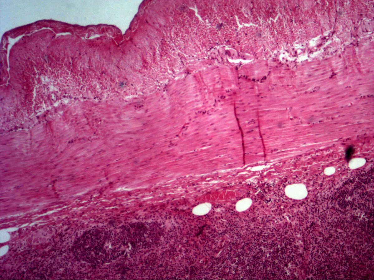
Labelled structures
This image has no labels – use the caption instead.
The tunica muscularis of the ileum is the classic specimen for smooth muscle: two layers of spindle-shaped muscle cells, an inner circular and an outer longitudinal one. In a transverse section of the gut one layer is therefore cut longitudinally and the other transversely – ideal for comparing both views side by side.
How to recognise it
- Spindle-shaped, mononuclear cells
- Centrally placed, elongated-oval nucleus
- Homogeneous eosinophilic cytoplasm without cross-striation
- In cross-section round profiles, many without a nucleus
- In the gut two layers: inner circular, outer longitudinal
Structures to find
- Circular layer of the tunica muscularis
- Longitudinal layer of the tunica muscularis
- Smooth muscle cells in longitudinal section with elongated nucleus
- Smooth muscle cells in cross-section
- Tunica serosa
- Mucosa with villi and crypts (for orientation)
Structure
Smooth muscle cells are mononuclear, spindle-shaped cells about 5–10 µm thick and up to several hundred µm long. The nucleus is central, located at the widest part of the cell, and is elongated-oval to cigar-shaped; in contracted cells it may look corkscrew-like. The cytoplasm is homogeneously eosinophilic because actin and myosin filaments are not arranged in sarcomeres – there is no cross-striation. Each cell is surrounded by a basal lamina and fine reticular fibres; cells are electrically coupled by gap junctions and form bundles separated by loose connective tissue.
Function and occurrence
Smooth muscle contracts slowly, persistently and economically and is controlled involuntarily by the autonomic nervous system, hormones and local stimuli. In the gut, the interplay of the circular layer (constriction) and the longitudinal layer (shortening) produces peristalsis; the myenteric (Auerbach) plexus between the two layers coordinates them. Other sites: walls of blood vessels, bronchi, ureter, urinary bladder, uterus, ductus deferens, arrector pili muscles and the intrinsic eye muscles.
Microscopy tips
In the overview, find the border between the two muscle layers. In longitudinal section you see parallel pink cells with elongated, centrally placed nuclei that are staggered relative to each other. In cross-section the cells appear as round profiles of varying size; only profiles cut through the middle of a cell contain a round nucleus, many are anucleate. Differential diagnosis: dense regular connective tissue (tendon) has thin, flat fibrocyte nuclei between thick collagen fibres and far fewer nuclei; skeletal and cardiac muscle show cross-striation.
Clinical relevance
Disorders of gut motility usually involve innervation, e.g. Hirschsprung disease, in which ganglion cells of the myenteric plexus are absent in the distal bowel. Benign smooth muscle tumours are called leiomyomas – very common in the uterus (fibroids); malignant variants are leiomyosarcomas. Under chronic load, such as outflow obstruction, smooth muscle responds with hypertrophy and hyperplasia – typically the thickened bladder wall in prostatic enlargement. In the gut, the muscle layer thickens proximal to a stenosis.
Slide images
Test your knowledge
Assign the correct number in the image to each term.
Tap the requested structure in the image.
Multiple-choice questions on structure and function.
Frequently asked questions
Where is the nucleus of a smooth muscle cell?
Centrally in the cell, at its widest part. It is elongated-oval and may appear wavy in contracted cells.
Why do many profiles lack a nucleus in cross-section?
The cells are spindle-shaped and staggered. A cross-section hits many cells at their thin ends, where there is no nucleus; only the thick mid-cell profiles show a round nucleus.
How do I tell smooth muscle from dense connective tissue?
Smooth muscle is more cellular, its nuclei are plumper, oval and lie within the cytoplasm. In dense connective tissue, few thin flat fibrocyte nuclei lie between thick, often wavy collagen fibres.
Related topics
Updated: