Skeletal muscle in cross-section and muscle spindle
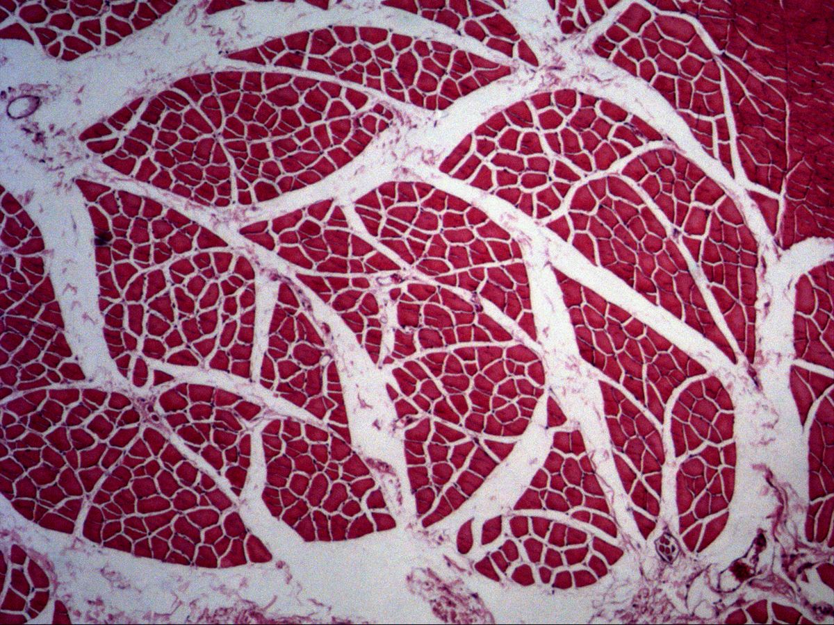
Masseter muscle · H&E
Labelled structures
This image has no labels – use the caption instead.
A cross-section of the masseter muscle shows the organisation of a skeletal muscle particularly well: muscle fibres are grouped into bundles separated by connective tissue septa. Between the bundles lie vessels, nerves and – as a small encapsulated sense organ – the muscle spindle.
How to recognise it
- Polygonal fibre profiles of similar size
- Nuclei peripheral beneath the sarcolemma
- Fine stippling of the sarcoplasm by myofibrils
- Fibre bundles separated by perimysium
- Muscle spindle: capsule with a few thin intrafusal fibres
Structures to find
- Skeletal muscle fibre in cross-section
- Peripheral nucleus
- Endomysium with capillaries
- Perimysium
- Muscle fibre bundle (fascicle)
- Muscle spindle with capsule and intrafusal fibres
Structure
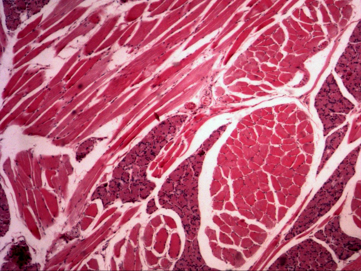
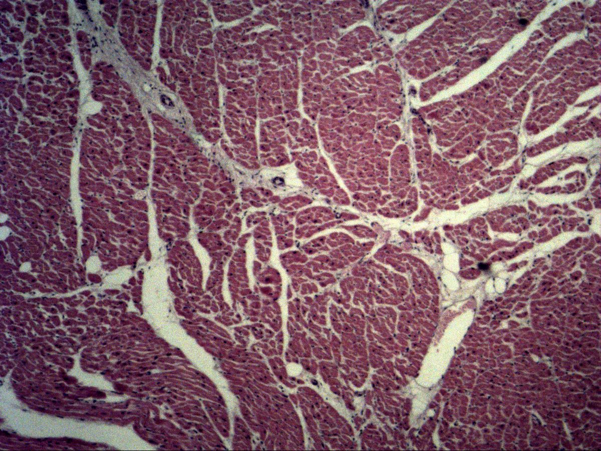
Skeletal muscle fibres in cross-section appear as polygonal, tightly packed profiles of similar size. The nuclei lie peripherally, directly beneath the sarcolemma; usually only one or two are seen per fibre profile. At high magnification the sarcoplasm shows a fine stippling from transversely cut myofibrils (Cohnheim fields). The endomysium surrounds every single fibre and carries capillaries, the perimysium groups fibres into bundles (fascicles) and carries larger vessels and nerves, and the epimysium covers the entire muscle.
Muscle spindle
The muscle spindle is a stretch receptor. It lies in the perimysium parallel to the working fibres and consists of a connective tissue capsule (a continuation of the perineurium), a fluid-filled space and a few distinctly thinner intrafusal muscle fibres (nuclear bag and nuclear chain fibres). These are wrapped by sensory Ia and II fibres and innervated by γ-motor neurons. The spindle signals muscle length and change in length to the spinal cord and underlies the stretch reflex, e.g. the jaw-jerk (masseter) reflex.
Microscopy tips
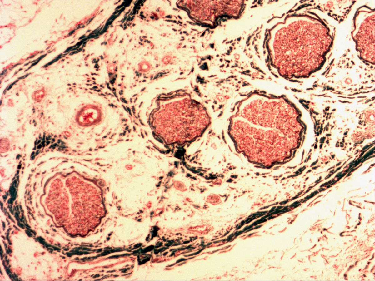
At low magnification, systematically scan the wider perimysial septa. A muscle spindle is a small round to oval structure with a thin, multilayered capsule containing a handful of thin muscle fibre profiles – often surrounded by a clear space. It can be confused with small nerve branches (many tiny axon profiles, perineurium, no eosinophilic muscle fibres) and blood vessels (endothelium, lumen, possibly erythrocytes). Wide clear gaps between fibres are usually shrinkage artefacts of fixation.
Clinical relevance
Muscle biopsies are mainly assessed in cross-section: variation in fibre size, central nuclei, fibre necrosis or grouped atrophic fibres help to distinguish myopathic from neurogenic disorders. An exaggerated jaw-jerk reflex indicates a lesion above the motor nucleus of the trigeminal nerve. Muscle spindles are clinically relevant too: their density is especially high in muscles for fine movements, such as hand and neck muscles. When testing stretch reflexes, a brief tap stretches the spindle and tests the reflex arc.
Slide images
Test your knowledge
Assign the correct number in the image to each term.
Tap the requested structure in the image.
Multiple-choice questions on structure and function.
Frequently asked questions
How do I recognise a muscle spindle?
By a thin connective tissue capsule in the perimysium enclosing a few much thinner muscle fibres. A clear space often surrounds the intrafusal fibres.
What is the difference between endomysium and perimysium?
The endomysium surrounds each muscle fibre and contains capillaries. The perimysium encloses whole fibre bundles and carries larger vessels and nerves.
Why don't I see a nucleus in every fibre?
The nuclei are spaced along the fibre, so a thin section only catches some of them.
Related topics
Updated: