Skeletal muscle
Tongue · H&E
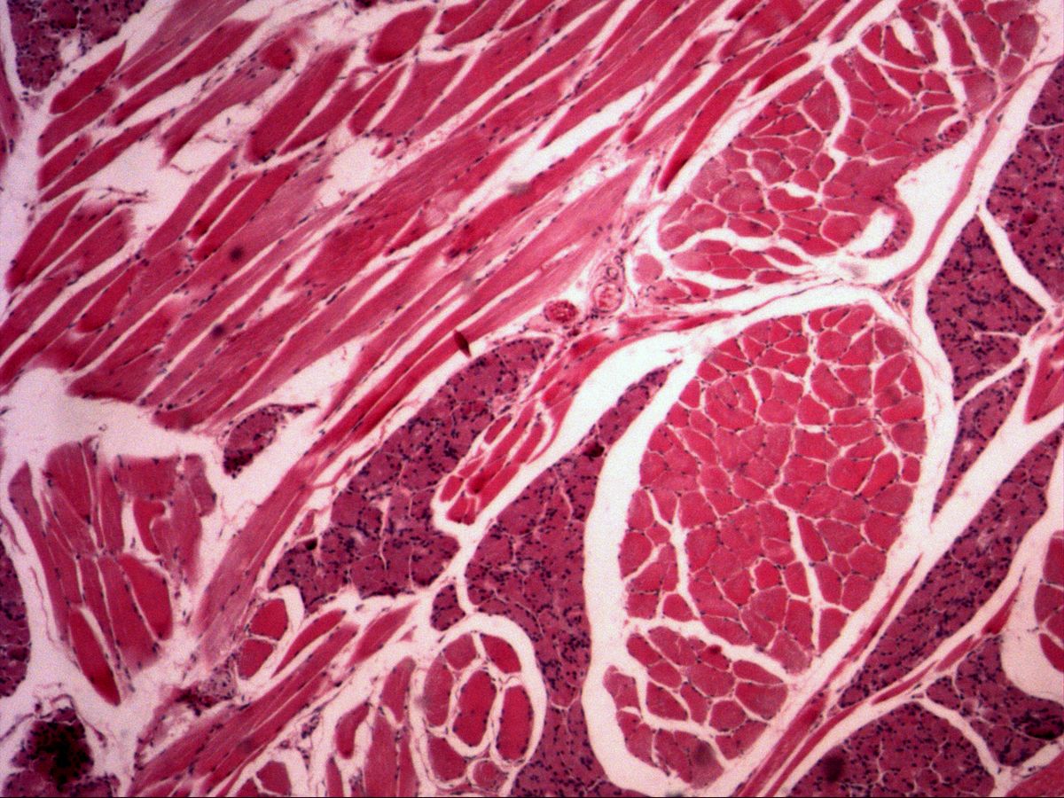
Labelled structures
This image has no labels – use the caption instead.
The tongue is an ideal place to study skeletal muscle: its intrinsic muscles run in three spatial directions, so the same section contains fibres cut longitudinally, transversely and obliquely. In longitudinal section, the cross-striation and the peripheral nuclei of the multinucleated muscle fibres stand out.
How to recognise it
- Long, cylindrical, unbranched fibres
- Distinct cross-striation (A and I bands)
- Many nuclei, located peripherally beneath the sarcolemma
- Endomysium between fibres, perimysium around bundles
- In the tongue, fibres run in three planes
Structures to find
- Longitudinally cut skeletal muscle fibre with cross-striation
- A-band (dark) and I-band (light)
- Peripheral nuclei
- Transversely cut muscle fibres
- Endomysium
- Perimysium
- Lingual glands among the muscle
Structure
A skeletal muscle fibre is a syncytium of fused myoblasts: up to several centimetres long, 10–100 µm thick, with hundreds of nuclei lying directly beneath the sarcolemma. The sarcoplasm is packed with myofibrils whose smallest contractile unit is the sarcomere between two Z-lines. The light I-bands (actin only) and dark A-bands (myosin, partly overlapping with actin) are in register across neighbouring myofibrils – producing the cross-striation seen in light microscopy. Connective tissue sheaths: endomysium around each fibre, perimysium around fibre bundles, epimysium around the whole muscle.
Function
Skeletal muscle contracts quickly and powerfully and is controlled voluntarily via motor end plates of α-motor neurons. During contraction, actin and myosin filaments slide past each other: the I-band and H-zone narrow while the A-band keeps its length. The tongue needs extremely fine changes of shape for speech, chewing and swallowing – made possible by the interweaving of longitudinal, transverse and vertical fibres.
Microscopy tips
In the overview, orient yourself by the differently cut fibre bundles and the pale connective tissue septa. To see the cross-striation you need high magnification and slight adjustment of the fine focus or a narrowed condenser diaphragm – light and dark bands then become clear. Check that the nuclei lie at the periphery of the fibre. Nuclei between fibres usually belong to fibrocytes, endothelial cells or satellite cells. Dark purple lobules among the muscle are lingual glands. Versus cardiac muscle: central nuclei, branching and intercalated discs there.
Clinical relevance
In muscular dystrophies (e.g. Duchenne) and myopathies nuclei move to a central position, fibre size varies greatly and fibres are replaced by fat and connective tissue – central nuclei in skeletal muscle fibres are therefore an important sign in muscle biopsies. Hypoglossal nerve palsy leads to atrophy of the ipsilateral tongue muscles; the protruded tongue deviates towards the affected side.
Slide images
Test your knowledge
Assign the correct number in the image to each term.
Tap the requested structure in the image.
Multiple-choice questions on structure and function.
Frequently asked questions
What causes the cross-striation?
By the regular arrangement of actin and myosin filaments in sarcomeres. Dark A-bands and light I-bands are in register across all myofibrils of a fibre.
Why are the nuclei at the periphery?
Myofibrils fill almost the entire sarcoplasm and push the nuclei beneath the sarcolemma. Central nuclei occur during regeneration and in muscle diseases.
Why is the tongue such a good specimen?
Because its intrinsic muscles interweave in three spatial directions. One section therefore shows longitudinally and transversely cut fibres side by side.
Related topics
Updated: