Adductor muscles of the hip

Thigh

Start quiz

This unit covers the muscles of the hip/femur, i.e. the adductor group: on the one hand adductor longus, brevis, magnus and minimus, and on the other hand obturator externus, pectineus and gracilis.

The adductors form the medial muscle group of the thigh. They arise from the pubis and ischium around the obturator foramen and mostly insert on the linea aspera of the femur; almost all are supplied by the obturator nerve. Besides adduction they stabilise the pelvis during standing and walking.

Key facts

  • The adductors arise from the pubis/ischium and mostly insert on the linea aspera.
  • The main nerve of the adductors is the obturator nerve (L2–L4).
  • Exceptions: posterior part of adductor magnus (tibial nerve) and pectineus (also femoral nerve).
  • Adductor magnus also inserts on the medial epicondyle; the adductor hiatus lies between its insertions.
  • Gracilis is the only two-joint adductor and inserts at the superficial pes anserinus.
  • Obturator externus passes below the femoral neck to the trochanteric fossa and rotates externally.

Adductor longus, brevis, magnus and minimus

Adductor longus

  • Origin: superior pubic ramus
  • Insertion: linea aspera (medial lip, middle third)

Adductor brevis

  • Origin: inferior pubic ramus
  • Insertion: linea aspera (upper third)

Adductor magnus / minimus (adductor minimus is the uppermost, often separate part of adductor magnus)

  • Origin: inferior pubic ramus; adductor magnus additionally from the ischial ramus and the ischial tuberosity
  • Insertion: linea aspera; the part of adductor magnus arising from the ischial tuberosity inserts by a tendon on the medial epicondyle of the femur (adductor tubercle)
  • Action (all): adduction (main action!), external rotation, pelvic stabilisation; adductor longus and brevis also flex, the posterior part of adductor magnus extends
  • Innervation: obturator nerve (L2–L4); the posterior, superficially placed (ischiocondylar) part of adductor magnus: tibial nerve (L4–L5)

Between the two insertions of adductor magnus lies the adductor hiatus, through which the femoral artery and vein reach the popliteal fossa. Adductor strains (especially of adductor longus) are typical sports injuries of the groin.

Obturator externus

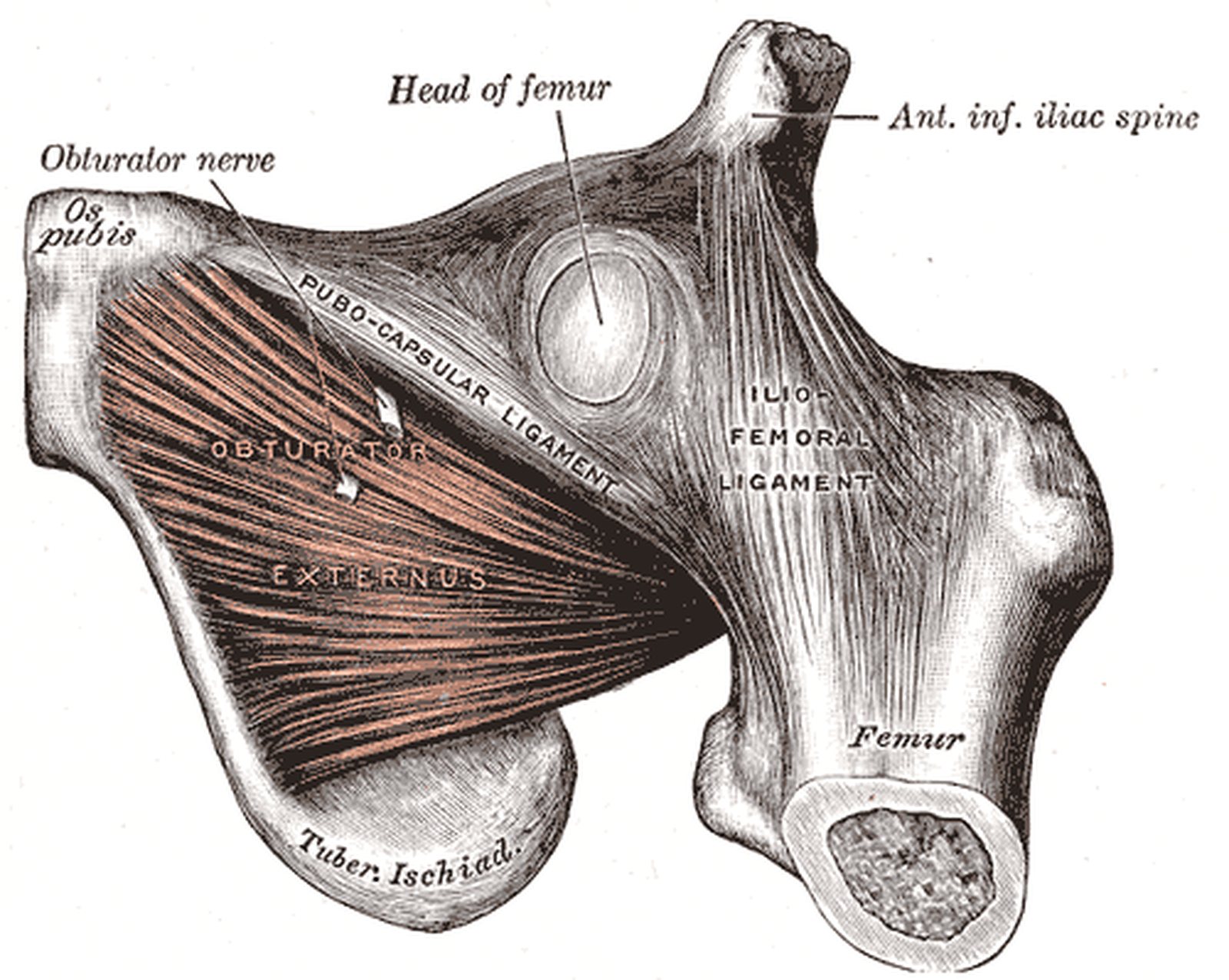
Hip from the front with obturator externus in reddish colour and the obturator nerve (Gray)
Illustration: Henry Gray, Anatomy of the Human Body (1918), public domain · Highlighting: AnatomyProf
  • Origin: outer surface of the obturator membrane and the bony margin of the obturator foramen
  • Insertion: trochanteric fossa
  • Action: adduction, external rotation
  • Innervation: obturator nerve (L2–L4)

The muscle lies deep to pectineus and the short adductors. Its tendon passes backwards below the femoral neck to the trochanteric fossa, so functionally it also belongs to the short external rotators of the hip. It is the only one of these rotators supplied by the obturator nerve, which reaches the thigh through the obturator canal along the upper border of the muscle. In Gray’s figure obturator externus is shown in reddish colour, with the obturator nerve labelled among its fibres; there is no yellow highlighting here.

Pectineus

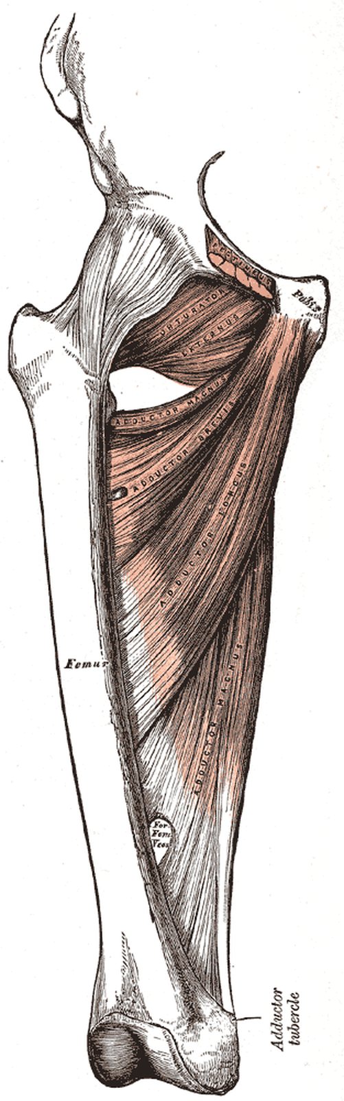
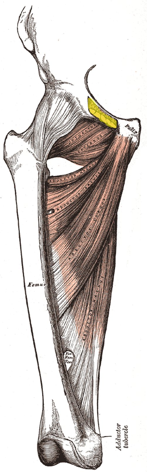
Thigh from the front with adductors; origin of pectineus on the pubis highlighted in yellow (Gray) Thigh from the front with adductors; origin of pectineus on the pubis highlighted in yellow (Gray)
Illustration: Henry Gray, Anatomy of the Human Body (1918), public domain · Highlighting: AnatomyProf
  • Origin: pecten pubis (pectineal line of the pubis)
  • Insertion: pectineal line of the femur (proximal continuation of the linea aspera)
  • Action: adduction, external rotation, flexion, pelvic stabilisation (frontal and sagittal planes)
  • Innervation: femoral nerve and obturator nerve (L2–L4) – dual innervation

Pectineus is a flat, quadrangular muscle at the junction between the anterior and medial thigh compartments. This intermediate position explains its dual innervation. Together with iliopsoas it forms the floor of the femoral triangle (iliopectineal fossa), in which the femoral artery and vein run. In the figure (thigh from the front, superficial muscles removed) the area of origin of pectineus on the pecten pubis is highlighted in yellow.

Gracilis

  • Origin: inferior pubic ramus (next to the symphysis)
  • Insertion: medial to the tibial tuberosity (superficial pes anserinus)
  • Action: hip: adduction, flexion; knee: flexion and internal rotation
  • Innervation: obturator nerve (L2–L4)

Gracilis is the most superficial and most medial adductor and the only one that crosses two joints, hip and knee. At the superficial pes anserinus it inserts together with sartorius and semitendinosus; beneath lies the anserine bursa, which can become painfully inflamed. Because it is long and functionally dispensable, its tendon is popular as a graft for anterior cruciate ligament reconstruction, and the muscle itself as a free muscle flap in plastic surgery.

Test your knowledge

Multiple-choice questions on structure and function.

Updated: