Ligaments of the ankle and foot

Ankle joints

Start quiz

This unit covers, among other things, the most important ligaments of the ankle joints. The tibiofibular ligaments, deltoid ligament, talofibular ligaments, calcaneofibular ligament and talocalcaneal ligaments are presented.

The upper ankle joint (talocrural joint) between the malleolar mortise and the talus is a hinge joint for dorsiflexion and plantar flexion; it is distinguished from the lower ankle joint (subtalar and talocalcaneonavicular joints), where pronation/eversion and supination/inversion take place. The syndesmosis holds the mortise together elastically, while the medial and lateral ligaments secure the joints at the sides.

Key facts

  • The anterior and posterior tibiofibular ligaments form the syndesmosis that holds the malleolar mortise together.
  • The deltoid (medial) ligament has four parts and limits pronation/eversion.
  • Lateral ligaments: anterior talofibular, calcaneofibular and posterior talofibular – they limit supination.
  • In an inversion sprain the anterior talofibular ligament tears first.
  • The interosseous talocalcaneal ligament separates the two chambers of the lower ankle joint.

Tibiofibular ligaments
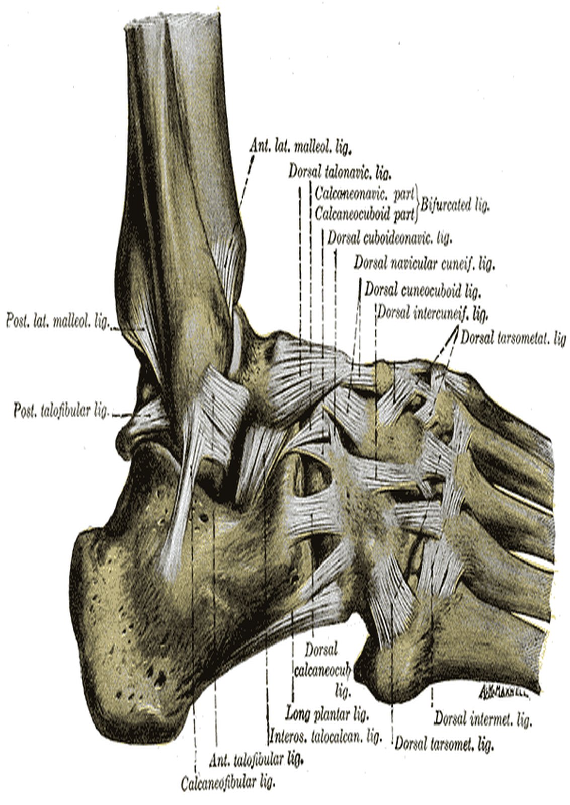
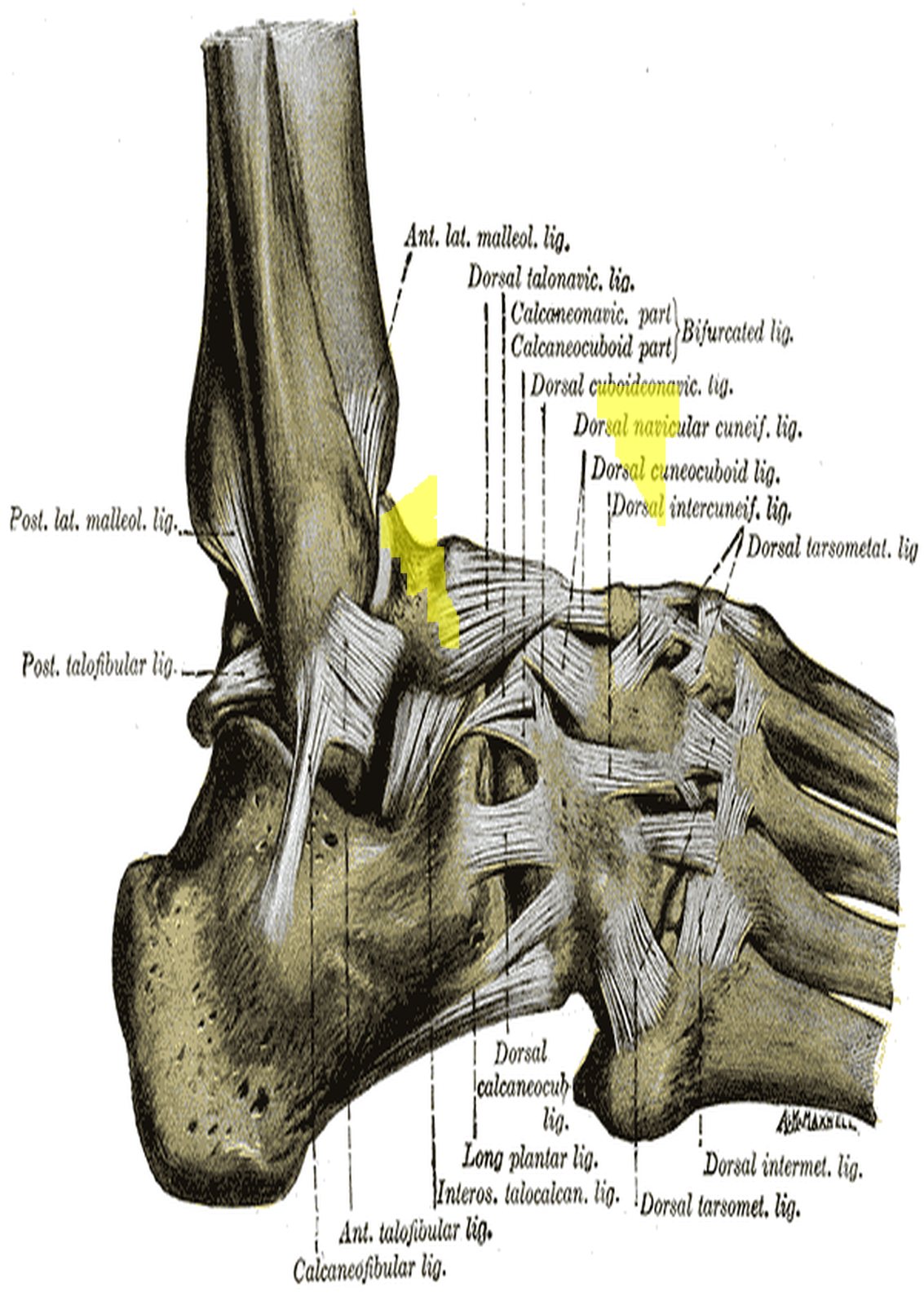
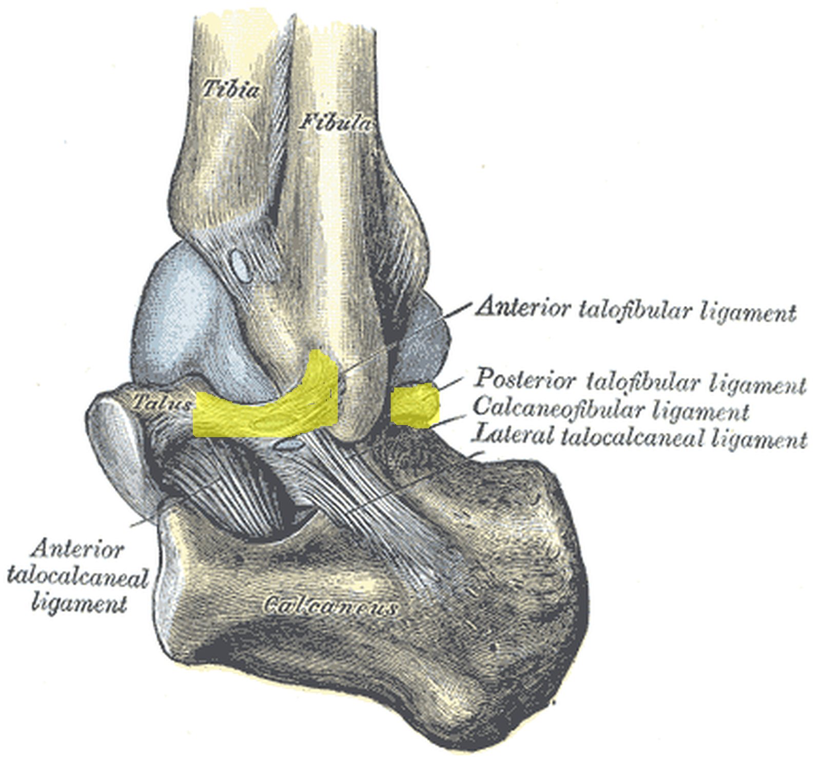
Foot and ankle from the lateral side; anterior tibiofibular ligament highlighted in yellow (Gray) Foot and ankle from the lateral side; anterior tibiofibular ligament highlighted in yellow (Gray)
Illustration: Henry Gray, Anatomy of the Human Body (1918), public domain · Highlighting: AnatomyProf
  • Anterior tibiofibular ligament: connects the anterolateral end of the tibia with the anterior border of the lateral malleolus of the fibula.
  • Posterior tibiofibular ligament: connects the posterolateral end of the tibia with the posterior border of the lateral malleolus.

Both ligaments form the tibiofibular syndesmosis; the interosseous membrane of the leg is another fibrous connection (syndesmosis) between tibia and fibula. The syndesmosis holds together the malleolar mortise that embraces the talus. Because the trochlea of the talus is wider anteriorly than posteriorly, the mortise widens slightly in dorsiflexion – the ligaments must therefore be strong yet elastic. In ankle fractures the syndesmosis is decisive for the Weber classification: in type C (fibular fracture above the syndesmosis) it is torn. In the figure (lateral view) the anterior tibiofibular ligament is highlighted in yellow; Gray labels it “Ant. lat. malleol. lig.”.

Deltoid ligament

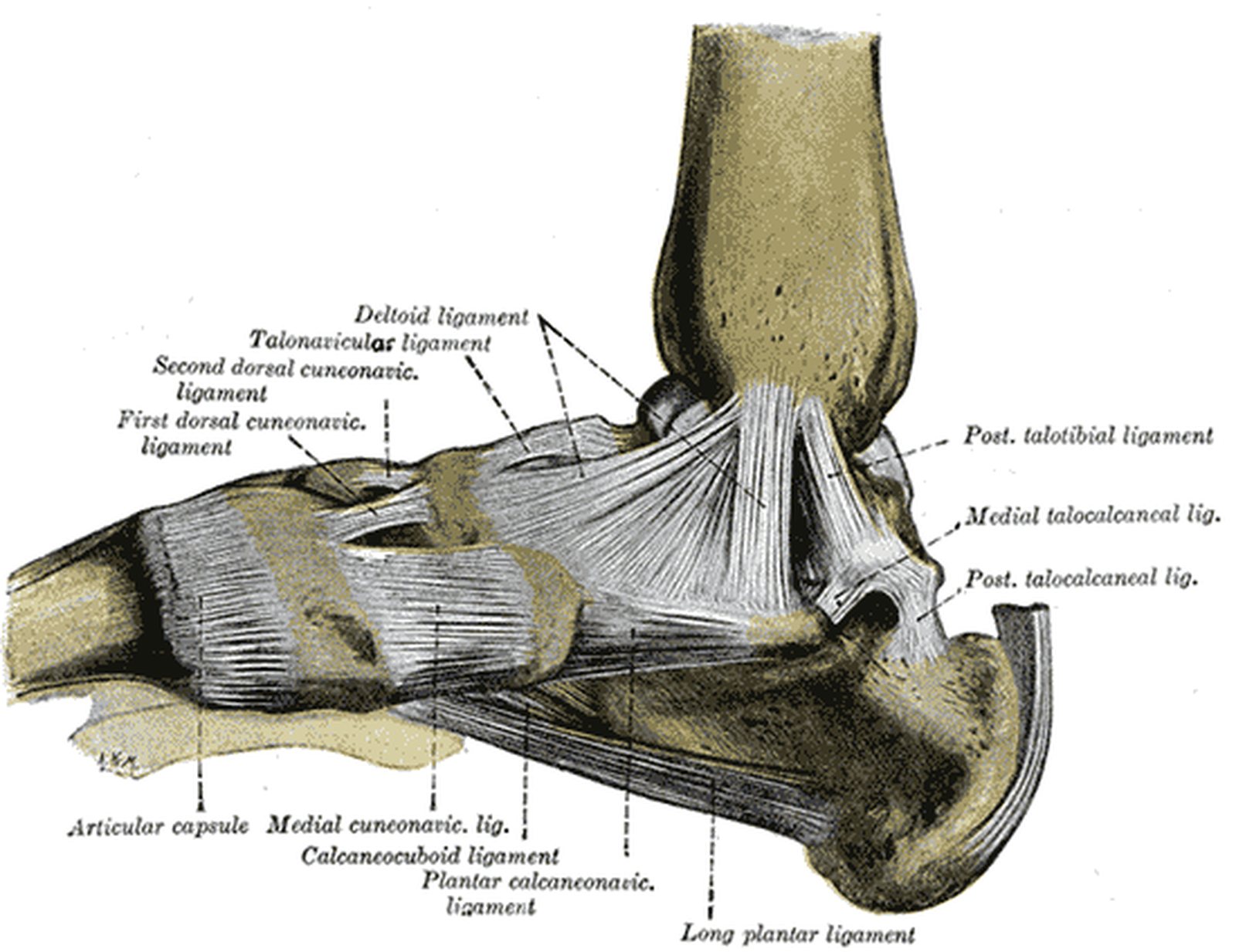
Foot from the medial side; tibiocalcaneal part of the deltoid ligament highlighted in yellow (Gray) Foot from the medial side; tibiocalcaneal part of the deltoid ligament highlighted in yellow (Gray)
Illustration: Henry Gray, Anatomy of the Human Body (1918), public domain · Highlighting: AnatomyProf

Also called the medial collateral ligament of the ankle, the deltoid ligament begins at the medial malleolus and fans out into four parts:

  • Anterior tibiotalar part – to the neck of the talus
  • Posterior tibiotalar part – to the posterior process of the talus
  • Tibiocalcaneal part – to the sustentaculum tali of the calcaneus
  • Tibionavicular part – to the tuberosity of the navicular

It is a very strong ligament that limits pronation or eversion. Because it spans both joints, it secures the upper ankle joint and – with its tibiocalcaneal and tibionavicular parts – also the lower ankle joint. Isolated ruptures are rare; in pronation injuries the medial malleolus tends to avulse instead. In the figure (medial view) the part running vertically from the medial malleolus to the calcaneus is highlighted in yellow, i.e. the tibiocalcaneal part inserting on the sustentaculum tali.

Talofibular ligaments

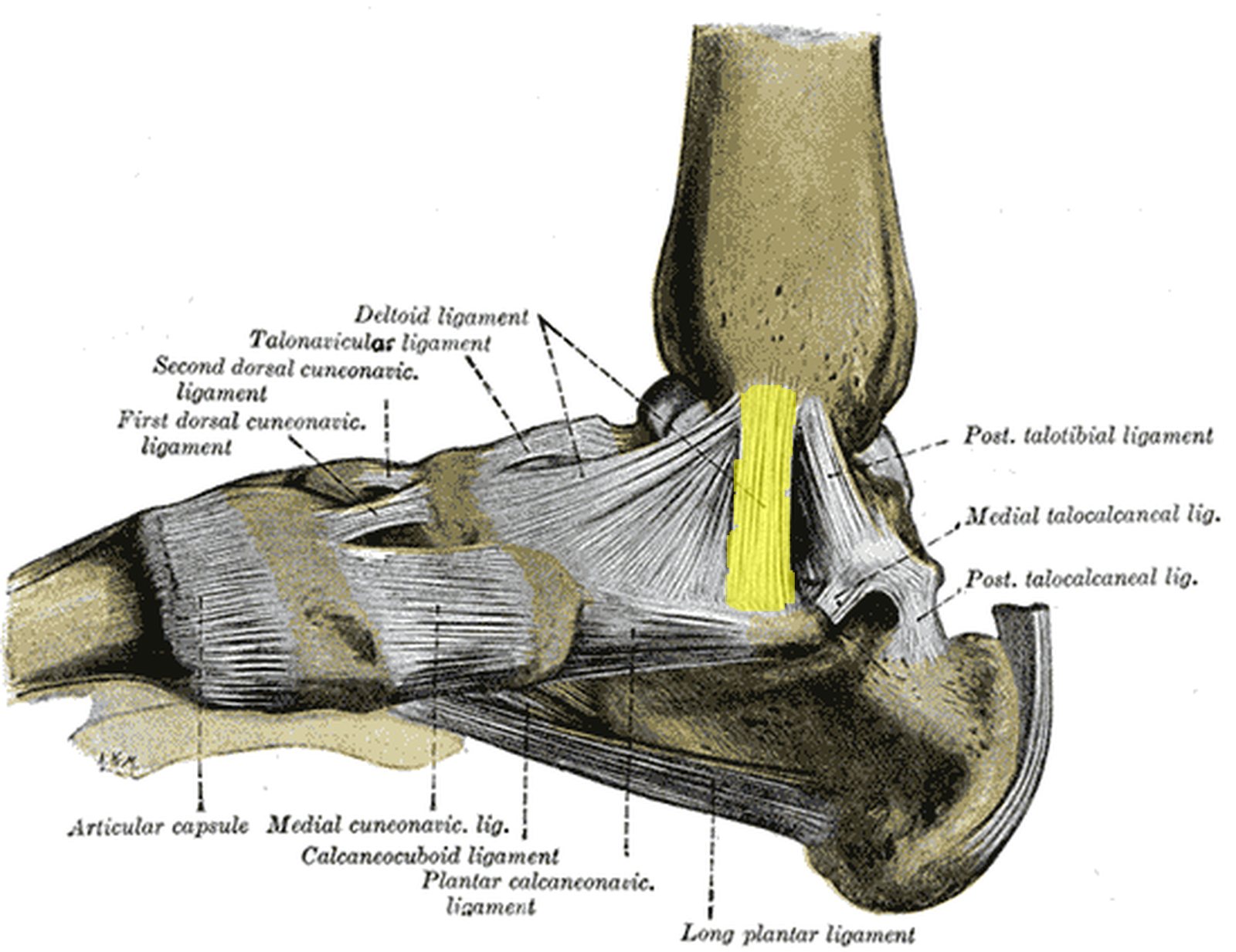
Ankle from posterolateral; anterior and posterior talofibular ligaments highlighted in yellow (Gray) Ankle from posterolateral; anterior and posterior talofibular ligaments highlighted in yellow (Gray)
Illustration: Henry Gray, Anatomy of the Human Body (1918), public domain · Highlighting: AnatomyProf

The two talofibular ligaments belong to the lateral ligament complex; both are attached to the lateral malleolus.

  • Anterior talofibular ligament: runs forwards to the neck of the talus.
  • Posterior talofibular ligament: runs backwards to the posterior process of the talus.

The anterior ligament is the weakest lateral ligament and is the first to be injured in the common inversion sprain (rolling the ankle outwards in plantar flexion); next the calcaneofibular ligament tears and – only in severe trauma – the strong posterior ligament. Instability of the anterior ligament is shown by the anterior drawer test of the ankle. In the figure (ankle from posterolateral) the anterior talofibular ligament between the lateral malleolus and the talar neck and the posterior talofibular ligament are highlighted in yellow.

Calcaneofibular ligament

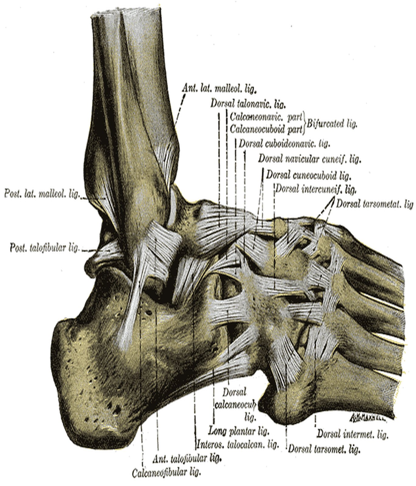
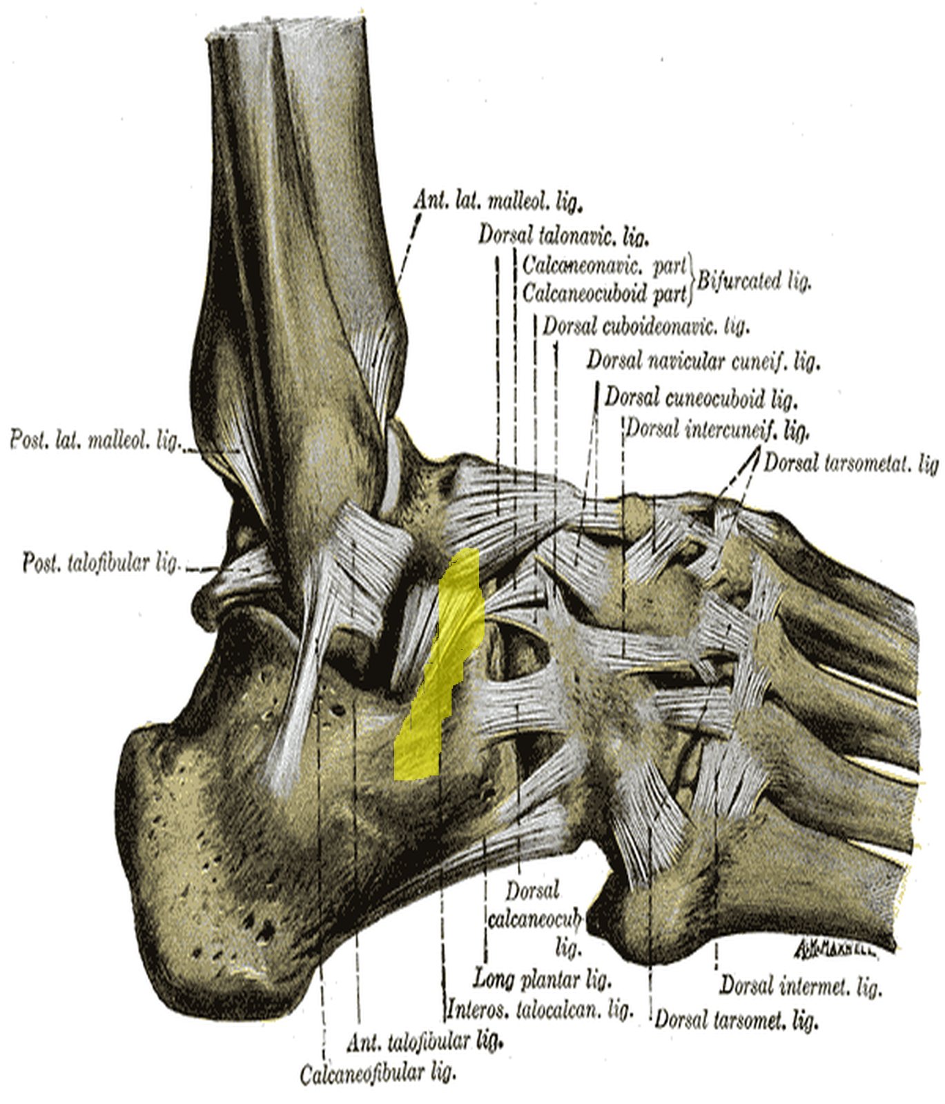
Foot from the lateral side with a ligamentous band on the lateral calcaneus highlighted in yellow (Gray) Foot from the lateral side with a ligamentous band on the lateral calcaneus highlighted in yellow (Gray)
Illustration: Henry Gray, Anatomy of the Human Body (1918), public domain · Highlighting: AnatomyProf
  • Course: the calcaneofibular ligament runs from the tip of the lateral malleolus backwards and downwards to a small tubercle on the lateral surface of the calcaneus.
  • Function: limits supination or inversion, especially in dorsiflexion.

It is the only one of the three lateral ligaments that spans both the upper and the lower ankle joint, stabilising both. It lies deep to the tendons of fibularis (peroneus) longus and brevis. In an inversion sprain it typically tears after the anterior talofibular ligament; a combined rupture is more likely to lead to chronic instability. Diagnosis is based on clinical findings, the talar tilt test and imaging if needed. In the figure (lateral view) a ligamentous band on the lateral side of the calcaneus below the ankle region is highlighted in yellow; Gray’s label “Calcaneofibular lig.” names the ligament.

Talocalcaneal ligaments

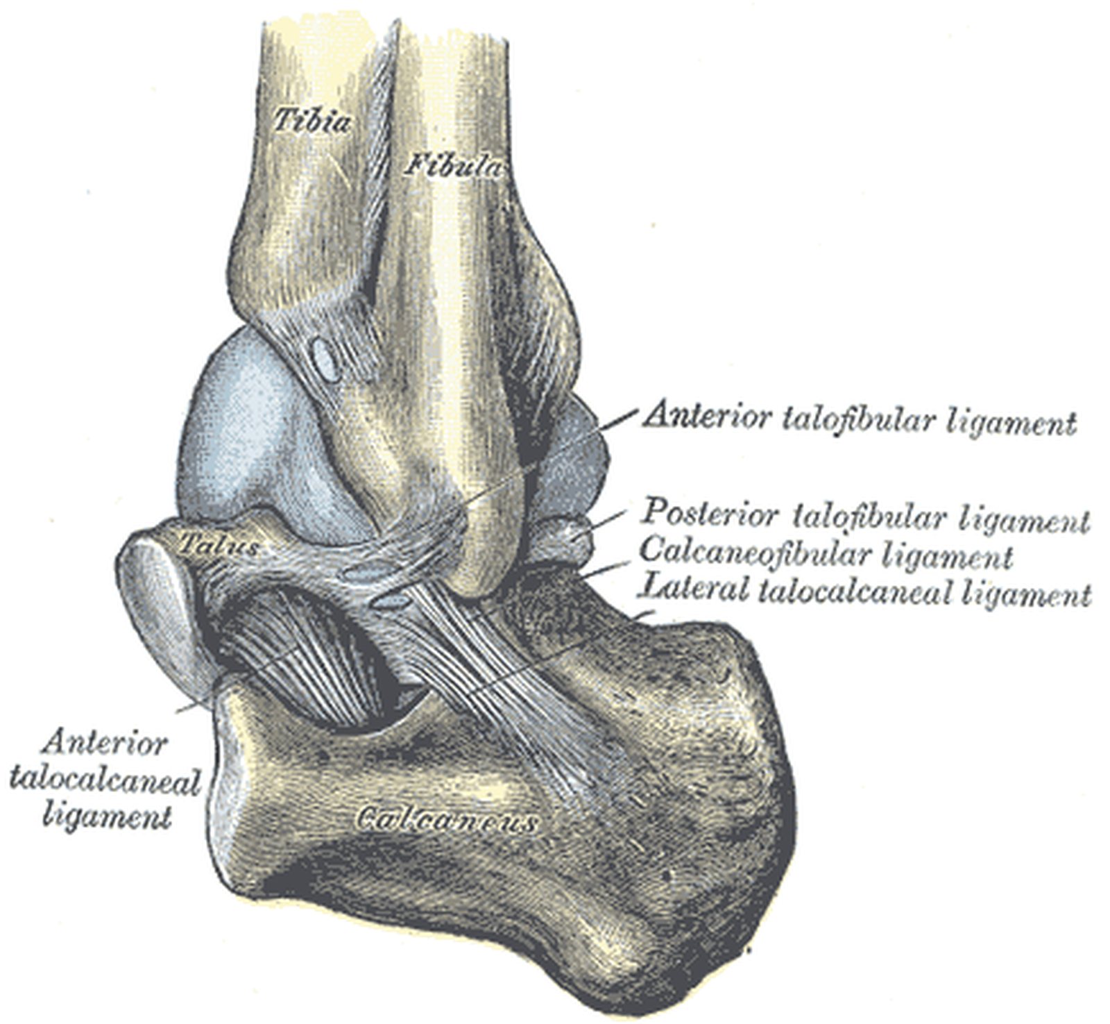
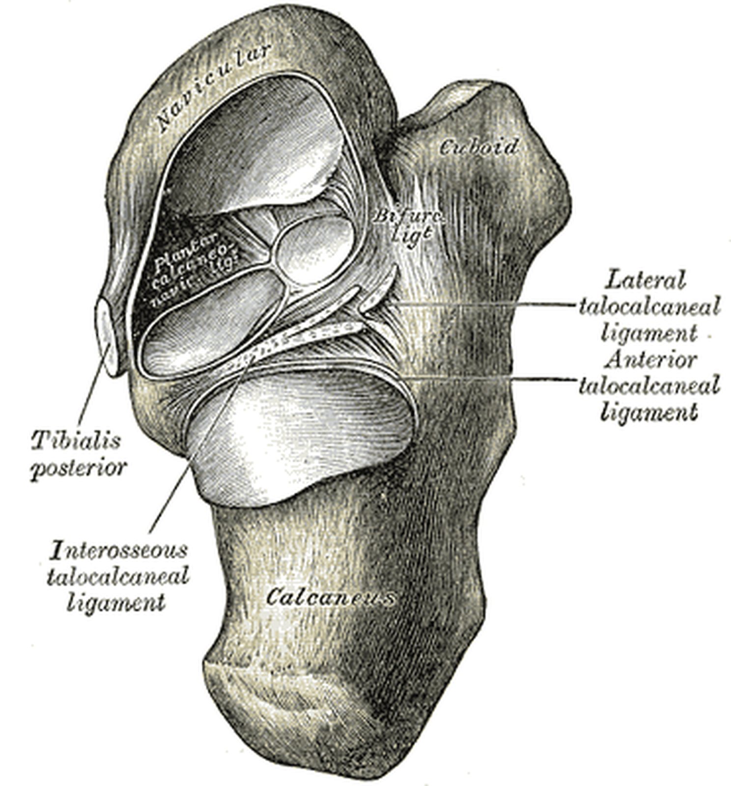
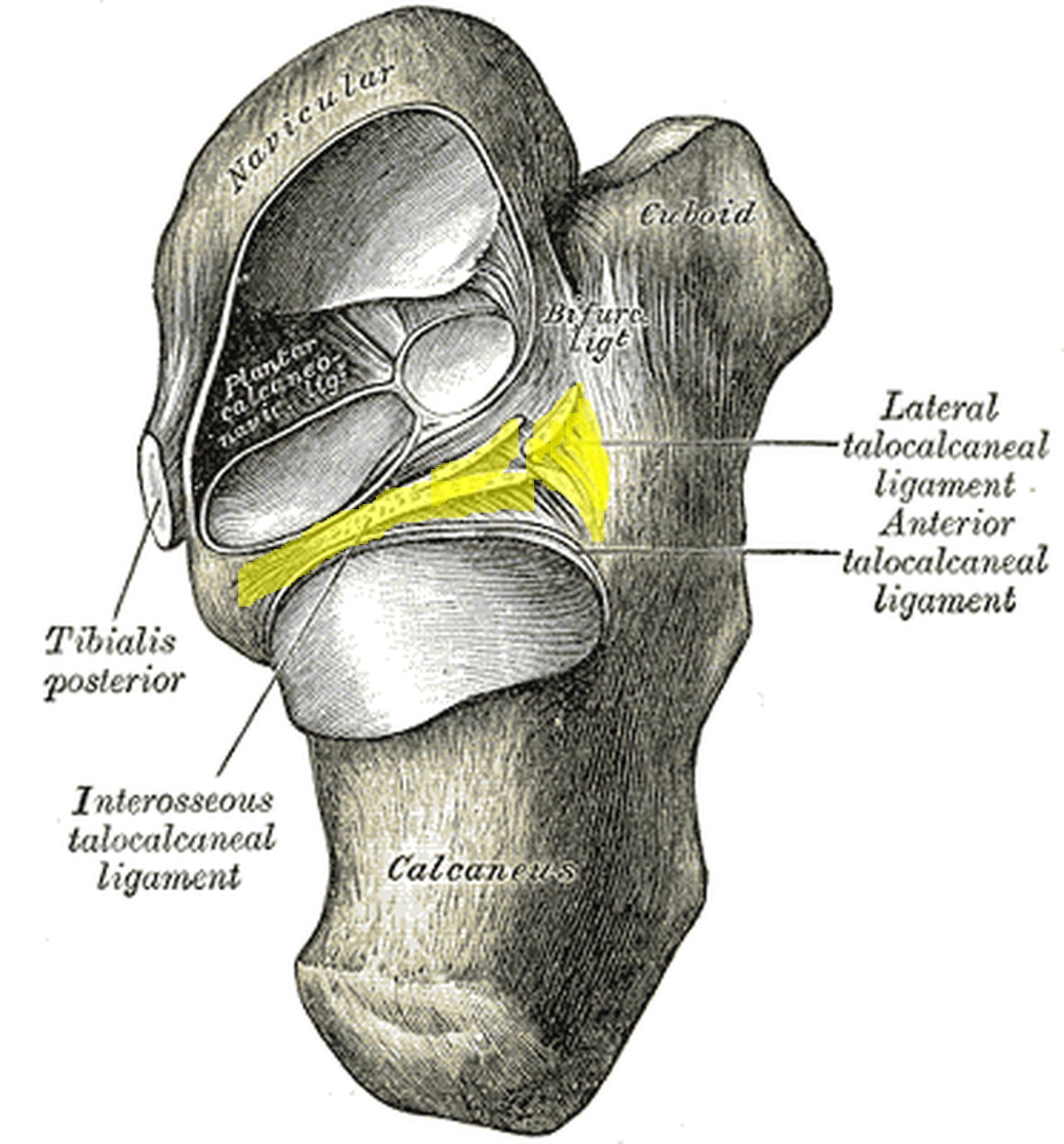
Calcaneus from above with the talus removed; interosseous and lateral talocalcaneal ligaments in yellow (Gray) Calcaneus from above with the talus removed; interosseous and lateral talocalcaneal ligaments in yellow (Gray)
Illustration: Henry Gray, Anatomy of the Human Body (1918), public domain · Highlighting: AnatomyProf
  • Interosseous talocalcaneal ligament: connects the heel bone (calcaneus) with the ankle bone (talus). It lies in the tarsal sinus/canal and separates the posterior chamber (subtalar joint) from the anterior chamber (talocalcaneonavicular joint) of the lower ankle joint.
  • Lateral talocalcaneal ligament: lies further out on the lateral side and limits supination.

As a rule, the medial ligaments limit pronation and the lateral ligaments limit supination. The strong interosseous talocalcaneal ligament is the most important ligament of the lower ankle joint; it lies close to the axis of movement and binds talus and calcaneus together in all positions. The medial and posterior talocalcaneal ligaments provide further reinforcement. In the figure (calcaneus from above after removal of the talus) the cut edges of the interosseous talocalcaneal ligament in the groove between the articular facets and the lateral talocalcaneal ligament are highlighted in yellow.

Test your knowledge

Multiple-choice questions on structure and function.

Updated: