Bones of the orbit
Orbit
This unit deals with the bones that form the orbit, making the structure of the eye socket clear. On the one hand, the participating bones – frontal, lacrimal, ethmoid, maxilla, sphenoid, palatine and zygomatic – are identified. On the other hand, structures such as the fissures and foramina are explained.
The orbit protects the eyeball together with its muscles, nerves and vessels. Through its openings it communicates with the middle cranial fossa, the pterygopalatine fossa, the nasal cavity and the face. Knowing which bones form each wall and what passes through which opening makes it possible to interpret fractures and nerve deficits in the orbital region.
Key facts
- Seven bones form the orbit: frontal, ethmoid, sphenoid, lacrimal, zygomatic, maxilla and palatine.
- The orbit is cone-shaped: base = orbital opening, apex = common tendinous ring.
- Optic canal: optic nerve and ophthalmic artery.
- Superior orbital fissure: cranial nerves III, IV, VI, branches of V1 and the superior ophthalmic vein.
- The lamina papyracea and the orbital floor are particularly thin – the floor is the typical site of a blow-out fracture.
Bones of the orbit
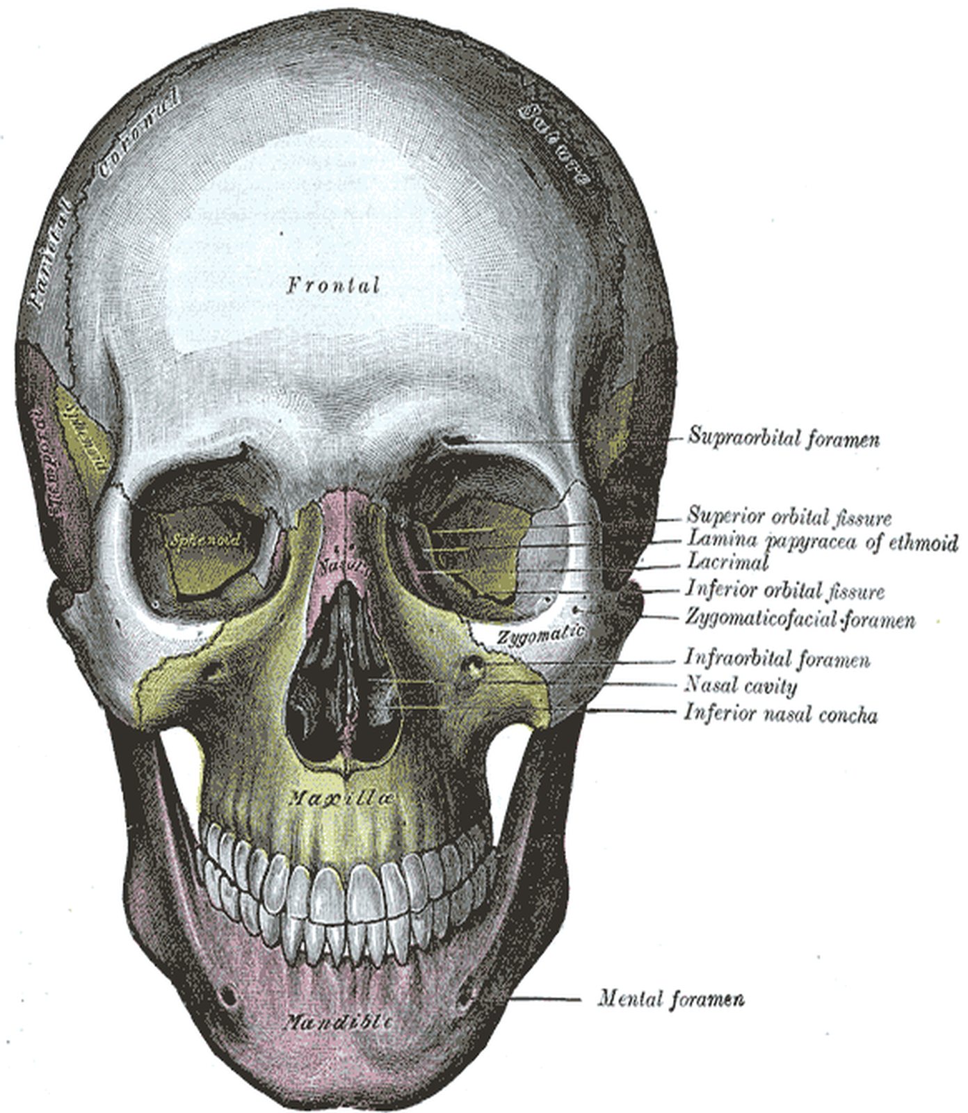
Structures of the orbit
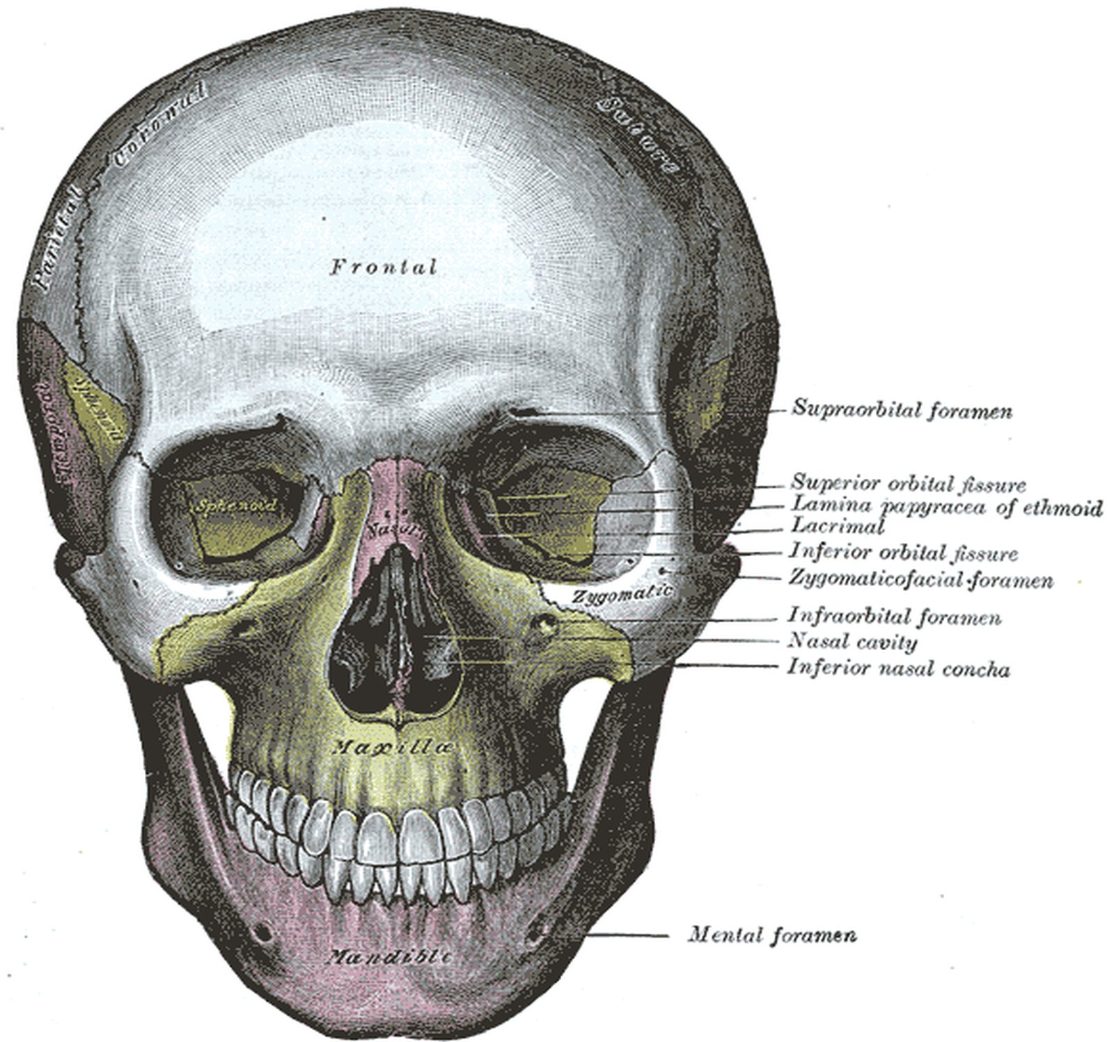
Test your knowledge
Multiple-choice questions on structure and function.
Frequently asked questions
Which bones form the orbit?
Seven bones: frontal, ethmoid, sphenoid, lacrimal, zygomatic, maxilla and palatine. The palatine bone contributes only a small orbital process to the orbital floor.
What is a blow-out fracture?
A fracture of the thin orbital floor (less often the medial wall) caused by a sudden rise in intraorbital pressure, e.g. from a fist or a ball. Tissue and the inferior rectus may herniate into the maxillary sinus; typical signs are double vision on upward gaze and numbness in the territory of the infraorbital nerve.
What passes through the superior orbital fissure?
The oculomotor (III), trochlear (IV) and abducens (VI) nerves, the branches of the ophthalmic nerve (frontal, lacrimal, nasociliary) and the superior ophthalmic vein.
Related topics
Updated: