Lamellar bone
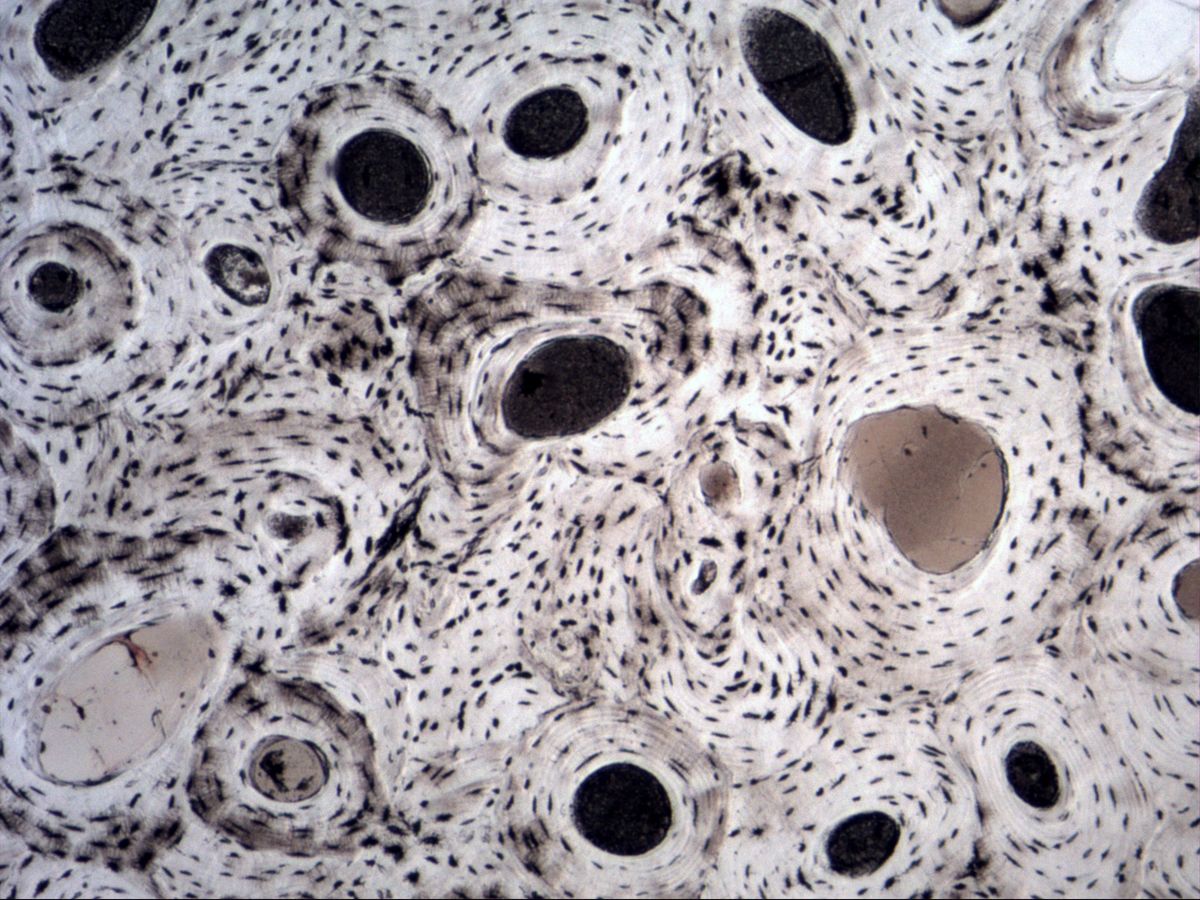
Tibia · ground section
Labelled structures
This image has no labels – use the caption instead.
A ground section is prepared by grinding dried, undecalcified bone until it is very thin. Organic components and cells are lost, but the cavities – canals, lacunae and canaliculi – are filled with air or grinding debris and appear dark. This makes the architecture of lamellar bone with its osteons particularly clear.
How to recognise it
- Rounded osteons with concentric lamellar rings
- Dark central Haversian canal in each osteon
- Spindle-shaped dark osteocyte lacunae between lamellae
- Fine, radiating canaliculi
- Interstitial lamellae as fragments between osteons
- No preserved cells (ground section)
Structures to find
- Osteon (Haversian system)
- Haversian canal
- Volkmann canal
- Concentric lamellae
- Interstitial lamellae
- Circumferential lamellae
- Osteocyte lacunae
- Canaliculi
Structure
The structural unit of compact bone is the osteon (Haversian system): a cylinder of 5–20 concentric lamellae around a central Haversian canal containing vessels and nerves. Transverse or oblique Volkmann canals connect Haversian canals with each other and with the periosteum and marrow cavity; they are not surrounded by their own lamellae.
Between osteons lie interstitial lamellae – remnants of older, partly resorbed osteons. Outer and inner circumferential lamellae close off the compact bone. Osteocytes lie in lacunae between the lamellae and are connected by fine canaliculi. Cement lines delimit each osteon.
Function
Within each lamella the type I collagen fibrils run parallel and helically, but at changing angles in neighbouring lamellae – similar to plywood. This makes lamellar bone very resistant to bending and torsion, while hydroxyapatite crystals provide compressive strength. The network of osteocytes and canaliculi serves for exchange of substances and for sensing mechanical load: osteocytes regulate the continuous remodelling by osteoclasts and osteoblasts via signalling molecules (e.g. sclerostin, RANKL). New osteons form when cutting cones of osteoclasts are refilled by osteoblasts.
Microscopy tips
In a ground section 'dark' means 'cavity': the large round black spots are Haversian canals, the small spindle-shaped black dots osteocyte lacunae. At high power you can see the hair-thin canaliculi radiating from them. Find an osteon with clearly visible rings and follow its border outwards – where lamellae end abruptly, the interstitial lamellae begin. Longitudinally or obliquely cut canals without their own lamellae suggest Volkmann canals. Compared with decalcified H&E sections: there the cells are preserved and the matrix is eosinophilic.
Clinical relevance
In osteoporosis resorption predominates: trabeculae become thinner and the compact bone porous, predisposing to fractures of vertebral bodies, femoral neck and distal radius. During fracture healing woven bone forms first and is subsequently remodelled into lamellar bone via osteon formation. In forensics, the density of osteons and interstitial lamellae can even give a rough estimate of age. In osteomyelitis, bacteria spread through the bone via Haversian and Volkmann canals.
Slide images
Test your knowledge
Assign the correct number in the image to each term.
Tap the requested structure in the image.
Multiple-choice questions on structure and function.
Frequently asked questions
Why are no cells visible in a ground section?
The bone is dried and ground, so the organic components are lost. Only the cavities in which the cells and vessels lay remain visible.
How do I tell Haversian and Volkmann canals apart?
Haversian canals run longitudinally in the centre of an osteon and are surrounded by concentric lamellae. Volkmann canals run transversely or obliquely, connect Haversian canals and pierce the lamellae without their own lamellar sheath.
What are interstitial lamellae?
Interstitial lamellae are remnants of older osteons that were partly resorbed during remodelling. They fill the gaps between intact osteons.
Related topics
Updated: