Intramembranous ossification
Fetal tissue · H&E
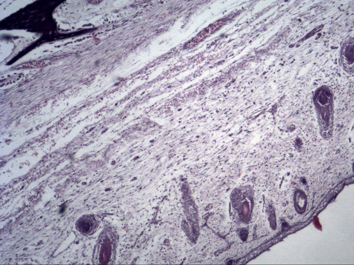
Labelled structures
This image has no labels – use the caption instead.
In intramembranous (direct) ossification bone forms directly from embryonic connective tissue, the mesenchyme, without a cartilage precursor. The flat bones of the skull vault, parts of the mandible and facial skeleton, and the clavicle develop this way. The images come from fetal head tissue and from the jaw region of a developing tooth.
How to recognise it
- Bone trabeculae directly within loose mesenchyme, no cartilage
- Osteoblast seam: row of dark cells on the trabecular surface
- Osteoid as a pale band between osteoblasts and mineralised matrix
- Osteocytes in numerous, irregularly distributed lacunae (woven bone)
- Occasional multinucleated osteoclasts in Howship's lacunae
Structures to find
- Mesenchyme
- Osteoblasts (osteoblast seam)
- Osteoid
- Osteocytes in lacunae
- Osteoclasts
- Howship's lacuna
- Bone trabeculae (woven bone)
Process
Mesenchymal cells condense in a well-vascularised area to form an ossification centre and differentiate into osteoblasts. These line up in an epithelium-like row (osteoblast seam) and secrete osteoid – the still uncalcified organic matrix of type I collagen. The osteoid then mineralises through deposition of hydroxyapatite.
Osteoblasts that wall themselves in become osteocytes in lacunae. This initially produces trabeculae of woven bone, which interconnect and are later remodelled into lamellar bone. The surrounding mesenchyme becomes periosteum or haematopoietic marrow.
Cells involved
- Osteoblasts: cuboidal to columnar, basophilic cytoplasm, eccentric nucleus; arranged in a row on the bone surface.
- Osteocytes: smaller, flattened cells in lacunae within the matrix, connected by processes.
- Osteoclasts: large, multinucleated, eosinophilic cells of the monocyte–macrophage lineage; lie in resorption bays (Howship's lacunae) and resorb bone.
- Mesenchymal cells: stellate cells in a loose, vascular ground substance around the trabeculae.
Microscopy tips
At low power first look for intensely stained, irregularly branching bone trabeculae within pale, loose mesenchyme – with no cartilage nearby. At high power examine the surface of the trabeculae: a row of closely packed dark cells is the osteoblast seam, a narrow pale band beneath it is osteoid. Inside, osteocytes lie in lacunae, much more irregularly arranged than in lamellar bone. Large multinucleated cells in bays are osteoclasts. Differential from endochondral ossification: there you find cartilage remnants and columnar cartilage.
Clinical relevance
In cleidocranial dysplasia (mutation of the transcription factor RUNX2, which controls osteoblast differentiation) intramembranous ossification is impaired: the clavicles are partly or completely absent, and fontanelles and sutures remain open for a long time. Adult fracture healing also proceeds partly by direct ossification when the fragments are rigidly fixed (primary bone healing). Premature closure of cranial sutures (craniosynostosis) leads to skull deformities.
Slide images
Test your knowledge
Assign the correct number in the image to each term.
Tap the requested structure in the image.
Multiple-choice questions on structure and function.
Frequently asked questions
Which bones form by intramembranous ossification?
Mainly the flat bones of the skull vault (frontal, parietal, parts of the occipital and temporal bones), much of the facial skeleton and mandible, and the clavicle, which forms by both intramembranous and endochondral ossification.
What is osteoid?
Osteoid is the not yet mineralised bone matrix of type I collagen and proteoglycans produced by osteoblasts. It appears as a narrow pale band between osteoblasts and calcified bone.
Does intramembranous ossification produce lamellar bone directly?
No. Woven bone with disorganised collagen fibres and many osteocytes forms first. It is later remodelled into lamellar bone.
Related topics
Updated: