Pharyngeal arches, pouches and clefts

Development of the pharynx

Start quiz

This unit deals with the developmental processes of the pharynx.

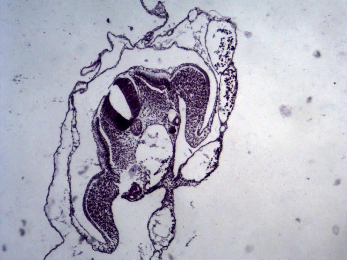
From the 4th embryonic week, the pharyngeal (branchial) arches appear in the head and neck region – swellings of mesoderm and migrating neural crest cells. Externally they are separated by ectodermal pharyngeal clefts (grooves), internally by endodermal pharyngeal pouches. Each arch receives its own nerve, artery, cartilage and musculature, which explains the innervation of many head and neck muscles in the adult.

Key facts

  • Pouches are endodermal, clefts are ectodermal; the arches contain mesoderm and neural crest cells.
  • 1st pouch: tympanic cavity and auditory tube; 1st cleft: external acoustic meatus.
  • 3rd pouch: thymus and inferior parathyroids; 4th pouch: superior parathyroids; 5th pouch: C cells.
  • Arch nerves: 1st mandibular, 2nd facial, 3rd glossopharyngeal, 4th and 6th vagus.
  • If the cervical sinus persists, lateral cervical cysts and fistulas develop.

Pharyngeal pouches
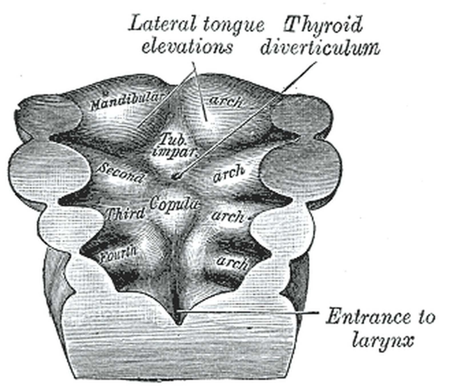
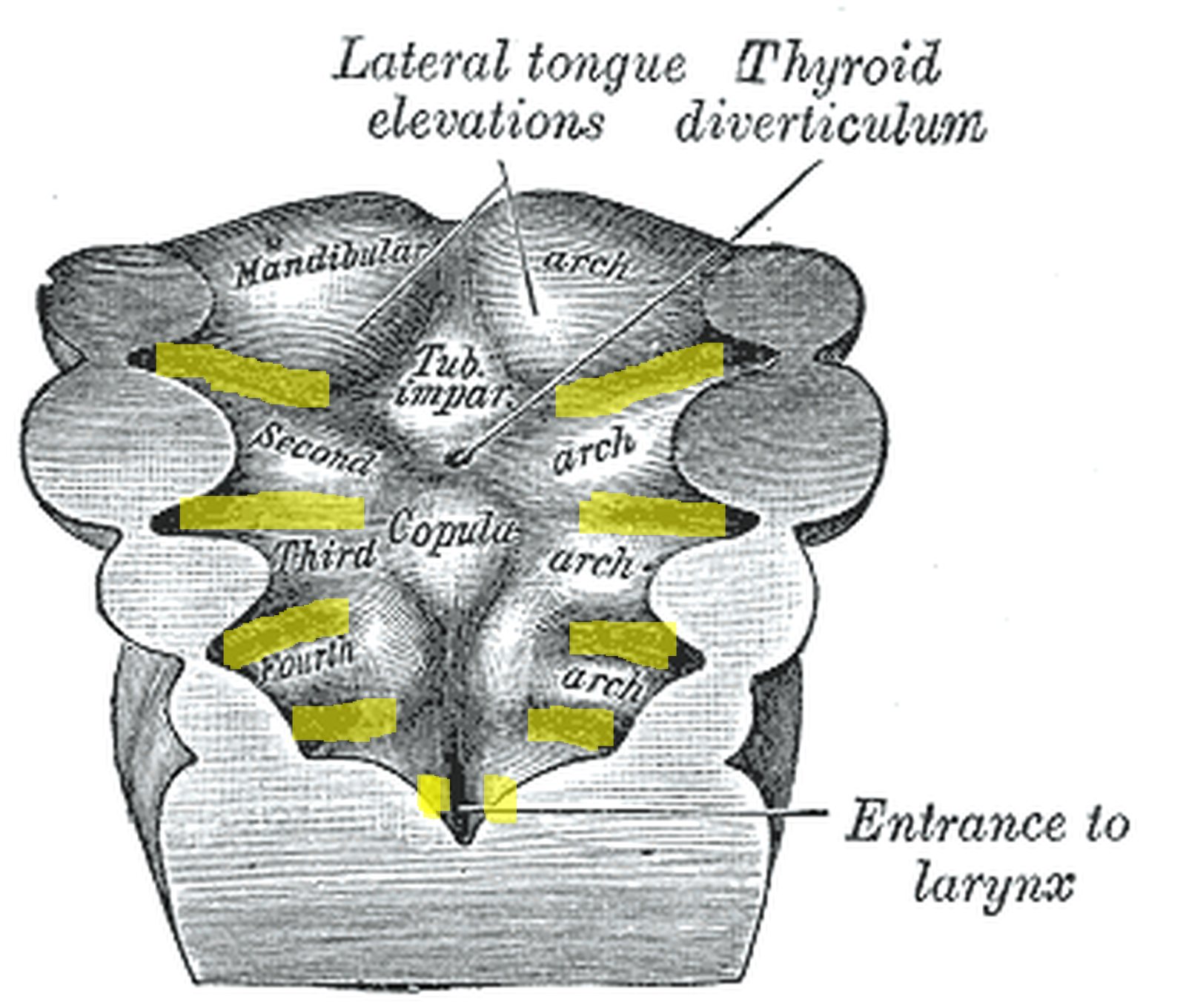
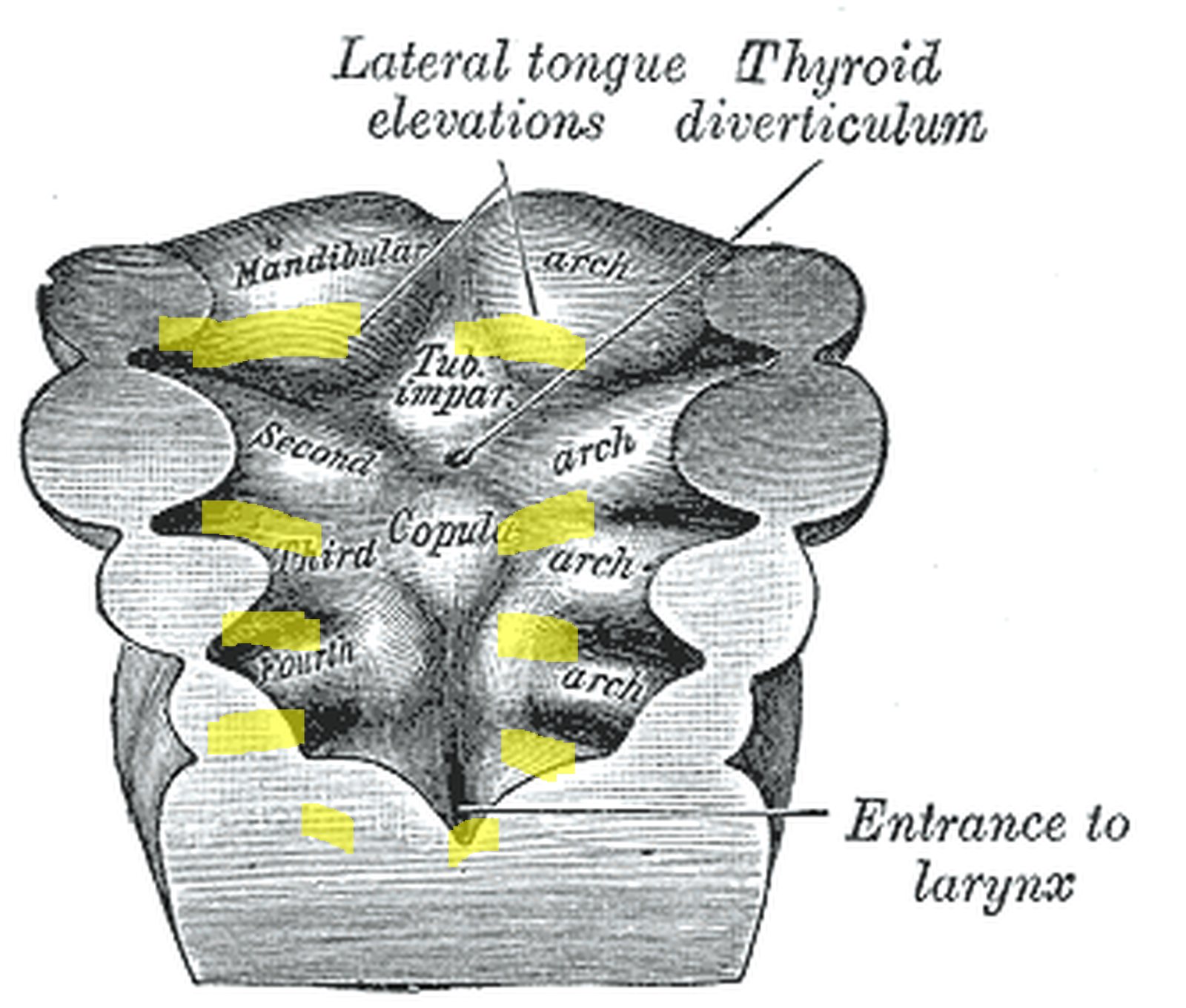
Floor of the embryonic pharynx with arches; pharyngeal pouches between the arches highlighted in yellow (Gray) Floor of the embryonic pharynx with arches; pharyngeal pouches between the arches highlighted in yellow (Gray)
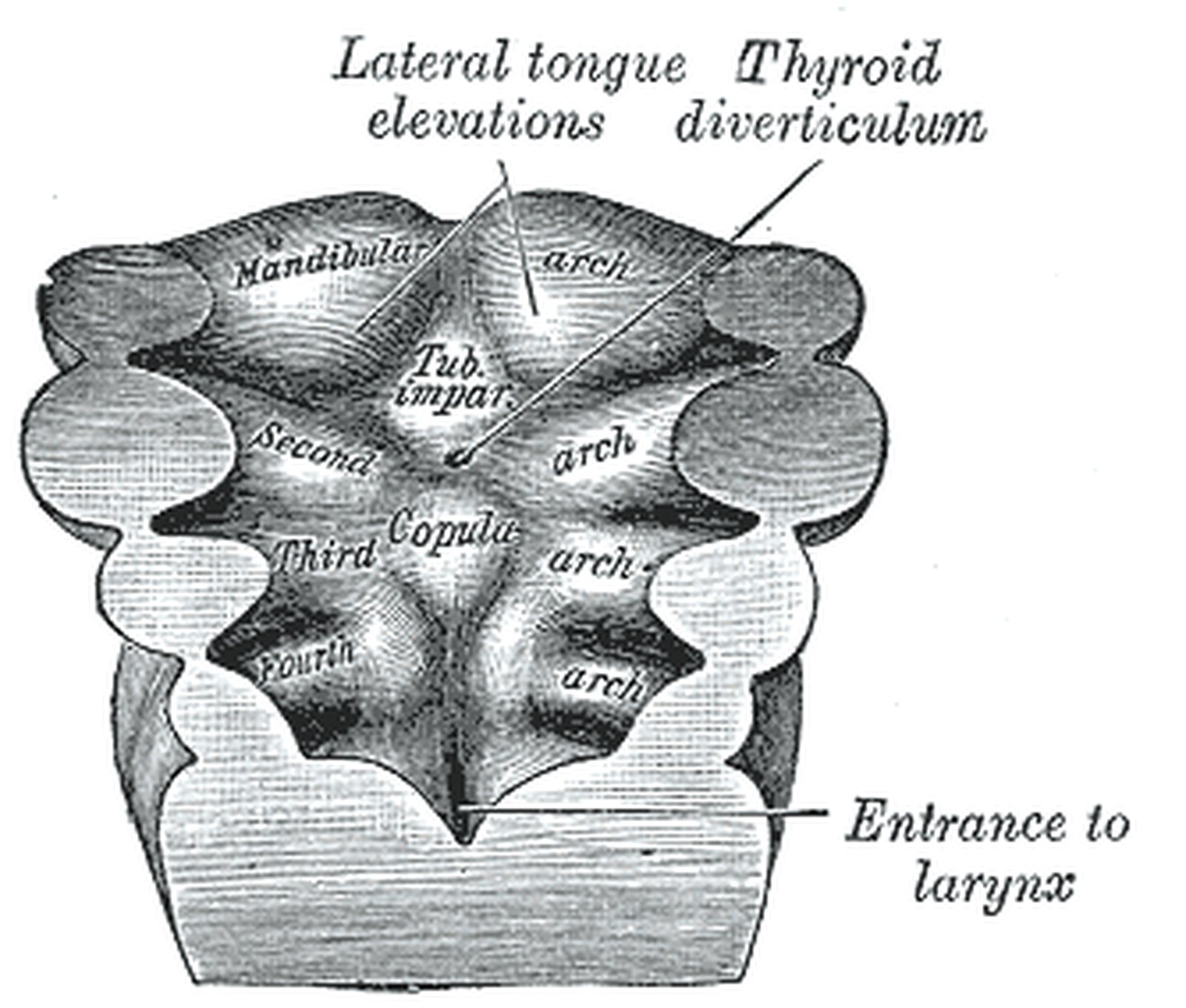
Illustration: Henry Gray, Anatomy of the Human Body (1918), public domain · Highlighting: AnatomyProf

The endodermal pharyngeal pouches are outpouchings of the foregut lying between the pharyngeal arches; five are counted, the fifth usually being rudimentary. They give rise to the following structures:

  • 1st pouch: tympanic cavity, auditory (Eustachian) tube
  • 2nd pouch: palatine tonsil, supratonsillar fossa
  • 3rd pouch: ventrally thymic epithelium, dorsally inferior parathyroid gland
  • 4th pouch: ventrally a small (mostly rudimentary) thymic contribution, dorsally superior parathyroid gland
  • 5th pouch: ultimobranchial body → C cells of the thyroid gland

The parathyroids from the 3rd pouch migrate caudally with the thymus and therefore end up below those from the 4th pouch. In DiGeorge syndrome (22q11 microdeletion) the 3rd and 4th pouches are affected: thymic hypoplasia with T-cell deficiency and hypocalcaemia due to absent parathyroids. In the figure (floor of the embryonic pharynx) the pouches between the arches are highlighted in yellow on both sides.

Pharyngeal clefts

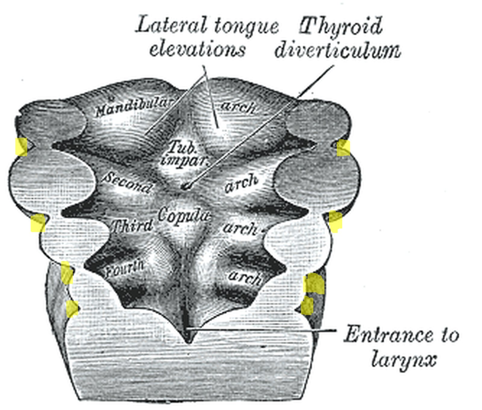
Embryonic pharyngeal arch region; clefts on the outer margin between the arches highlighted in yellow (Gray) Embryonic pharyngeal arch region; clefts on the outer margin between the arches highlighted in yellow (Gray)
Illustration: Henry Gray, Anatomy of the Human Body (1918), public domain · Highlighting: AnatomyProf

The pharyngeal clefts (grooves) are the external, ectodermal depressions between the pharyngeal arches:

  • 1st cleft: external acoustic meatus and outer (ectodermal) layer of the tympanic membrane.
  • 2nd, 3rd and 4th clefts: are overgrown by the rapidly growing 2nd arch and form the cervical sinus, which later regresses.

Note: if the cervical sinus fails to regress, lateral cervical (branchial) cysts or fistulas develop. They typically lie along the anterior border of the sternocleidomastoid; a complete fistula may extend to the tonsillar fossa (remnant of the 2nd pouch). Median neck cysts, by contrast, arise from remnants of the thyroglossal duct. The eardrum therefore has three layers: ectoderm outside (1st cleft), mesenchyme in the middle and endoderm inside (1st pouch). In the figure the notches between the arches on the outer margin – the pharyngeal clefts – are highlighted in yellow.

Pharyngeal arches

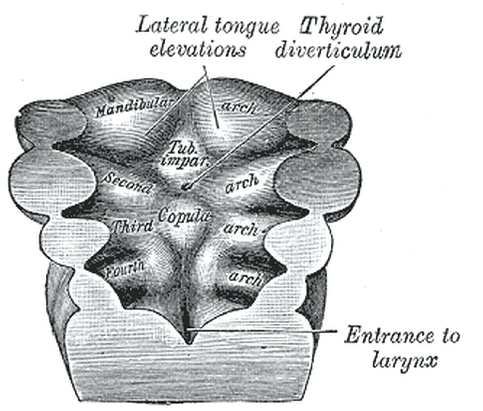
Floor of the embryonic pharynx; mandibular arch and 2nd to 4th pharyngeal arches highlighted in yellow (Gray) Floor of the embryonic pharynx; mandibular arch and 2nd to 4th pharyngeal arches highlighted in yellow (Gray)
Illustration: Henry Gray, Anatomy of the Human Body (1918), public domain · Highlighting: AnatomyProf

Each pharyngeal arch contains its own muscles, nerve and cartilage:

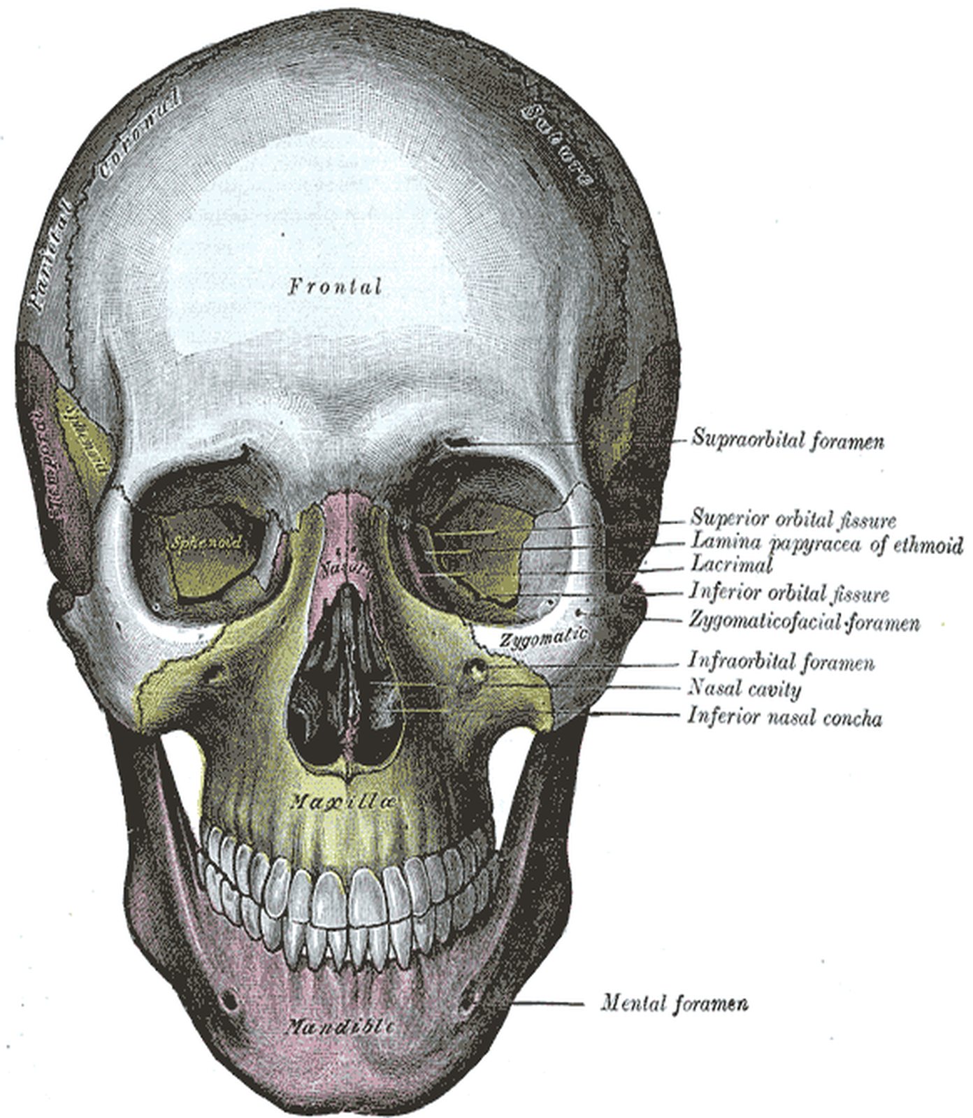
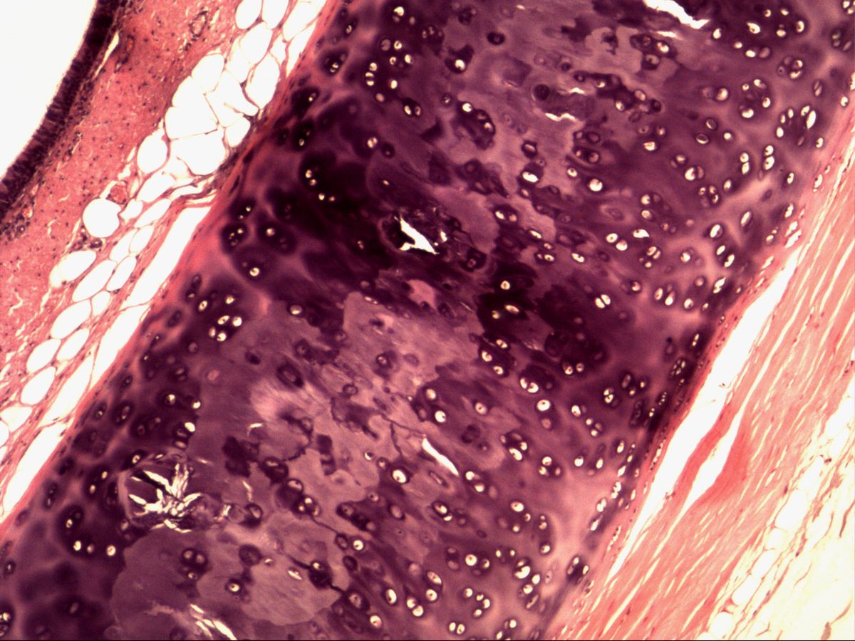
  • 1st arch (mandibular arch): muscles of mastication; mandibular nerve (V3); Meckel’s cartilage (→ malleus, incus)
  • 2nd arch (hyoid arch): muscles of facial expression; facial nerve; Reichert’s cartilage (→ stapes, styloid process, lesser horn of the hyoid)
  • 3rd arch: stylopharyngeus – some accounts also assign upper pharyngeal and soft palate muscles to it, which are now mostly attributed to the 4th arch; glossopharyngeal nerve; body (lower part) and greater horn of the hyoid bone
  • 4th arch: pharyngeal constrictors, soft palate muscles (except tensor veli palatini), cricothyroid; vagus nerve (superior laryngeal nerve); thyroid cartilage
  • 6th arch (the 5th is rudimentary in humans): intrinsic laryngeal muscles; vagus nerve (recurrent laryngeal nerve); cricoid and arytenoid cartilages

These assignments explain, for example, why tensor tympani (1st arch) is supplied by the trigeminal nerve and stapedius (2nd arch) by the facial nerve. In the figure the arches themselves are highlighted in yellow; Gray labels the “Mandibular arch” and the “Second”, “Third” and “Fourth arch”.

Test your knowledge

Multiple-choice questions on structure and function.

Updated: