Ligaments of the knee

Knee joint

Start quiz

This unit presents the ligaments of the knee. They stabilise the joint and also limit its movements (ligamentous restraint) in the knee joint (articulatio genus). The menisci, cruciate ligaments, patellar ligament, patellar retinaculum, collateral ligaments and popliteal ligaments are covered.

The knee is the largest joint of the human body and functionally a rotating hinge joint: it allows flexion and extension and, in the flexed position, rotation of the leg. Because the femoral condyles and the tibial plateau hardly fit each other, its stability depends largely on the ligaments and menisci – which is why these structures are so often injured in sport.

Key facts

  • The menisci compensate for the incongruence between femoral condyles and tibial plateau; the medial meniscus is fused with the MCL and injured more often.
  • ACL: lateral femoral condyle → anterior intercondylar area; PCL: medial femoral condyle → posterior intercondylar area.
  • The cruciate ligaments secure the joint mainly in flexion and check internal rotation; the collateral ligaments are taut in extension and check external rotation.
  • The fibular collateral ligament lies outside the capsule; the tibial collateral ligament is fused with the capsule and the medial meniscus.
  • The patellar ligament and the retinacula are part of the quadriceps extensor mechanism.
  • The oblique and arcuate popliteal ligaments reinforce the posterior capsule and prevent hyperextension.

Menisci
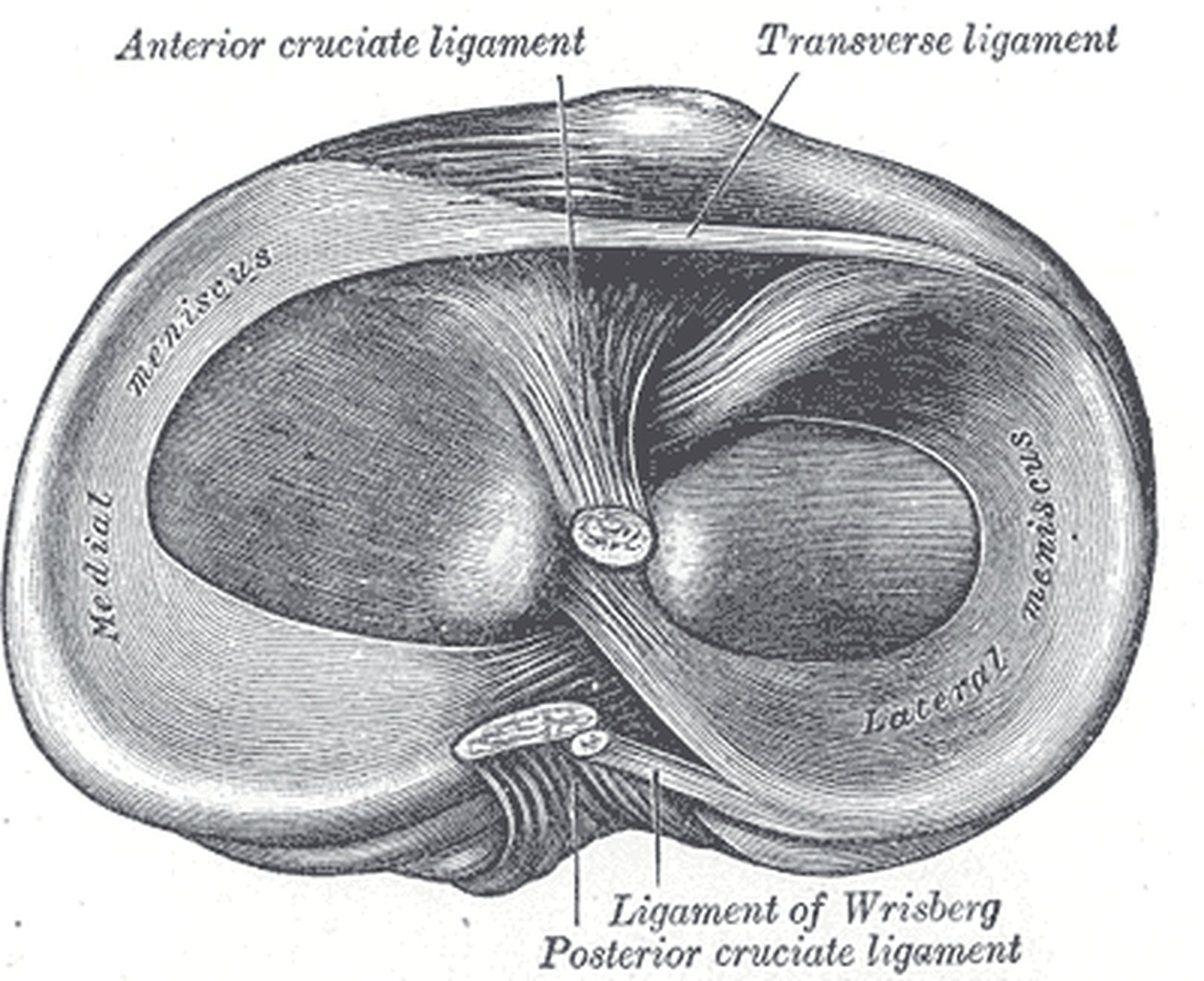
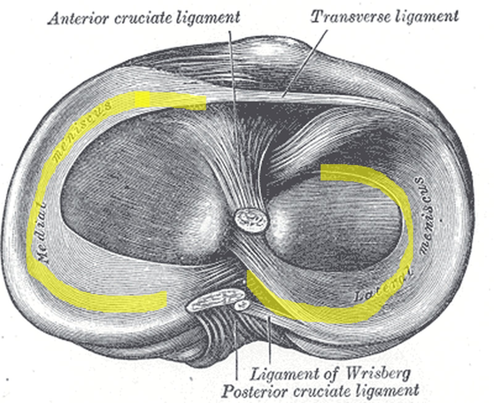
Head of the tibia from above with the medial and lateral menisci highlighted in yellow (Gray) Head of the tibia from above with the medial and lateral menisci highlighted in yellow (Gray)
Illustration: Henry Gray, Anatomy of the Human Body (1918), public domain · Highlighting: AnatomyProf

The two menisci (medial and lateral meniscus) are crescent-shaped discs of fibrocartilage and dense connective tissue with a wedge-shaped cross-section. Each meniscus is divided into three parts: anterior horn (cornu anterius), intermediate part (pars intermedia) and posterior horn (cornu posterius).

  • The anterior and posterior horns are attached to the intercondylar area of the tibia.
  • The anterior horns are joined by the transverse ligament of the knee.
  • Together the menisci form a congruent socket on the tibial condyles; during flexion (rolling movement) they glide backwards.

The menisci enlarge the contact area, distribute pressure and absorb shock; only their outer rim is vascularised, so inner tears heal poorly. The C-shaped medial meniscus is fused with the capsule and the medial collateral ligament, is therefore less mobile and is injured far more often than the almost circular lateral meniscus. In the figure (head of the tibia from above) the medial and lateral menisci are highlighted in yellow.

Cruciate ligaments

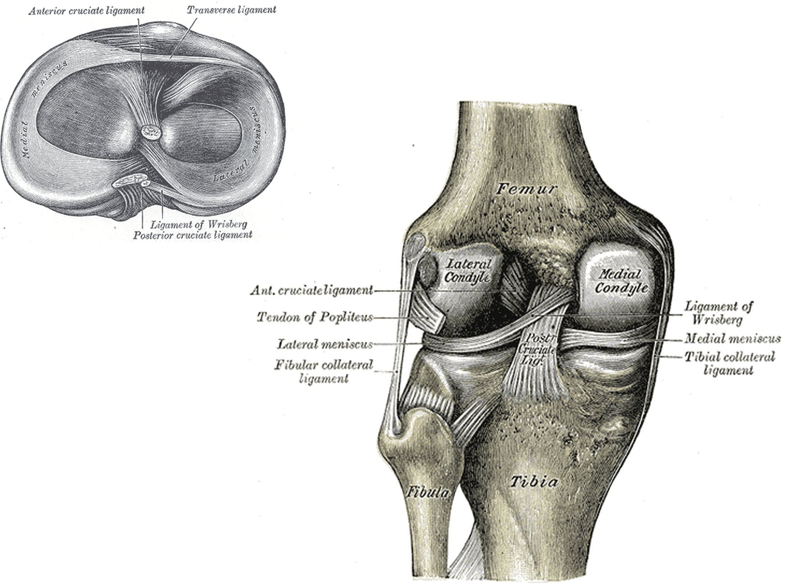
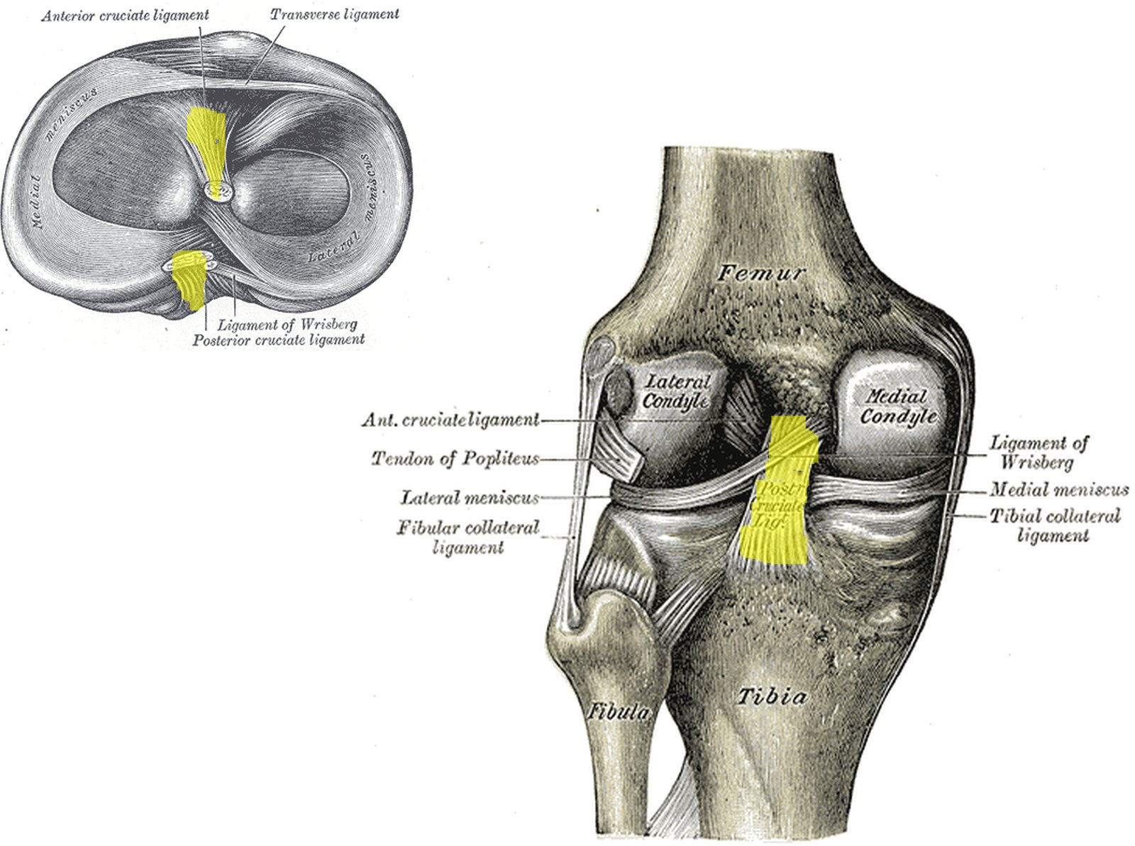
Tibial head from above and knee from behind; cruciate attachments and posterior cruciate ligament in yellow (Gray) Tibial head from above and knee from behind; cruciate attachments and posterior cruciate ligament in yellow (Gray)
Illustration: Henry Gray, Anatomy of the Human Body (1918), public domain · Highlighting: AnatomyProf

The cruciate ligaments lie in the intercondylar fossa – inside the fibrous layer of the joint capsule but outside the synovial membrane.

  • Anterior cruciate ligament (ACL): from the inner surface of the lateral femoral condyle to the anterior intercondylar area.
  • Posterior cruciate ligament (PCL): from the inner surface of the medial femoral condyle to the posterior intercondylar area.

They prevent anterior and posterior displacement of the tibia relative to the femur, secure the joint especially in flexion and check internal rotation, because they wind around each other. ACL rupture after twisting injuries (football, skiing) is common; it is tested with the Lachman test and the anterior drawer test. In the “unhappy triad” the ACL, the medial collateral ligament and the medial meniscus are injured together. In the figure the tibial attachments of both cruciate ligaments (left, tibia from above) and the posterior cruciate ligament (right, posterior view) are highlighted in yellow.

Patellar ligament

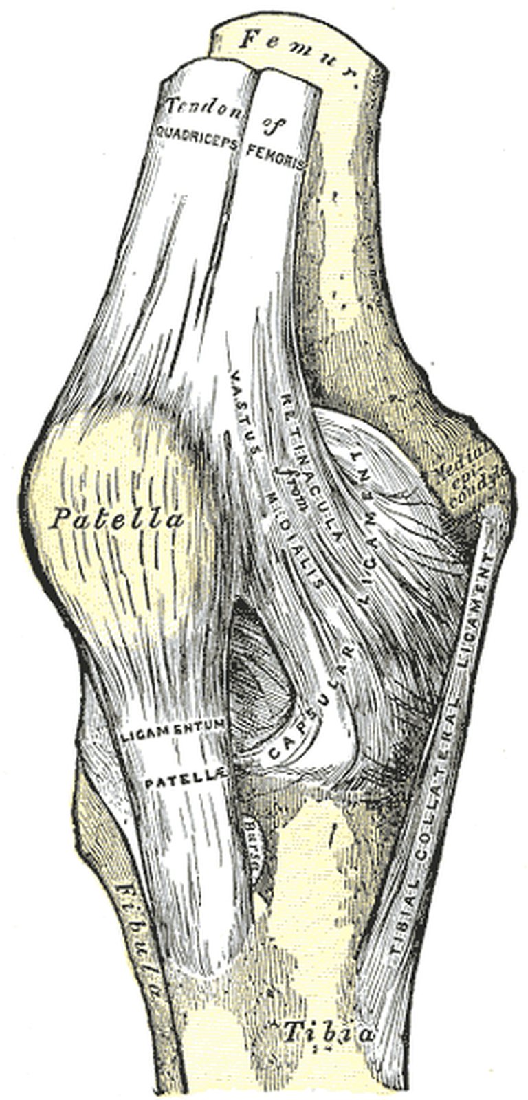
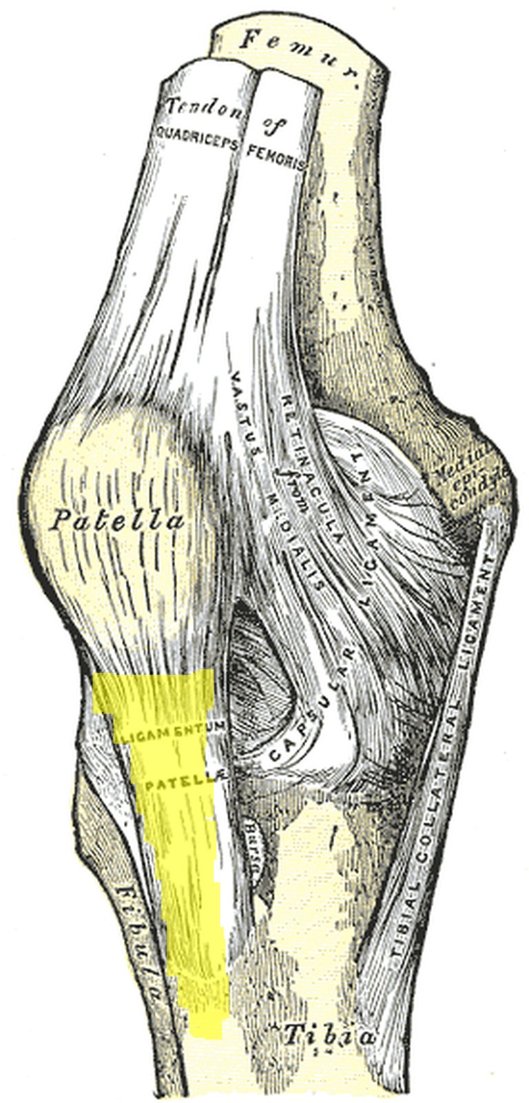
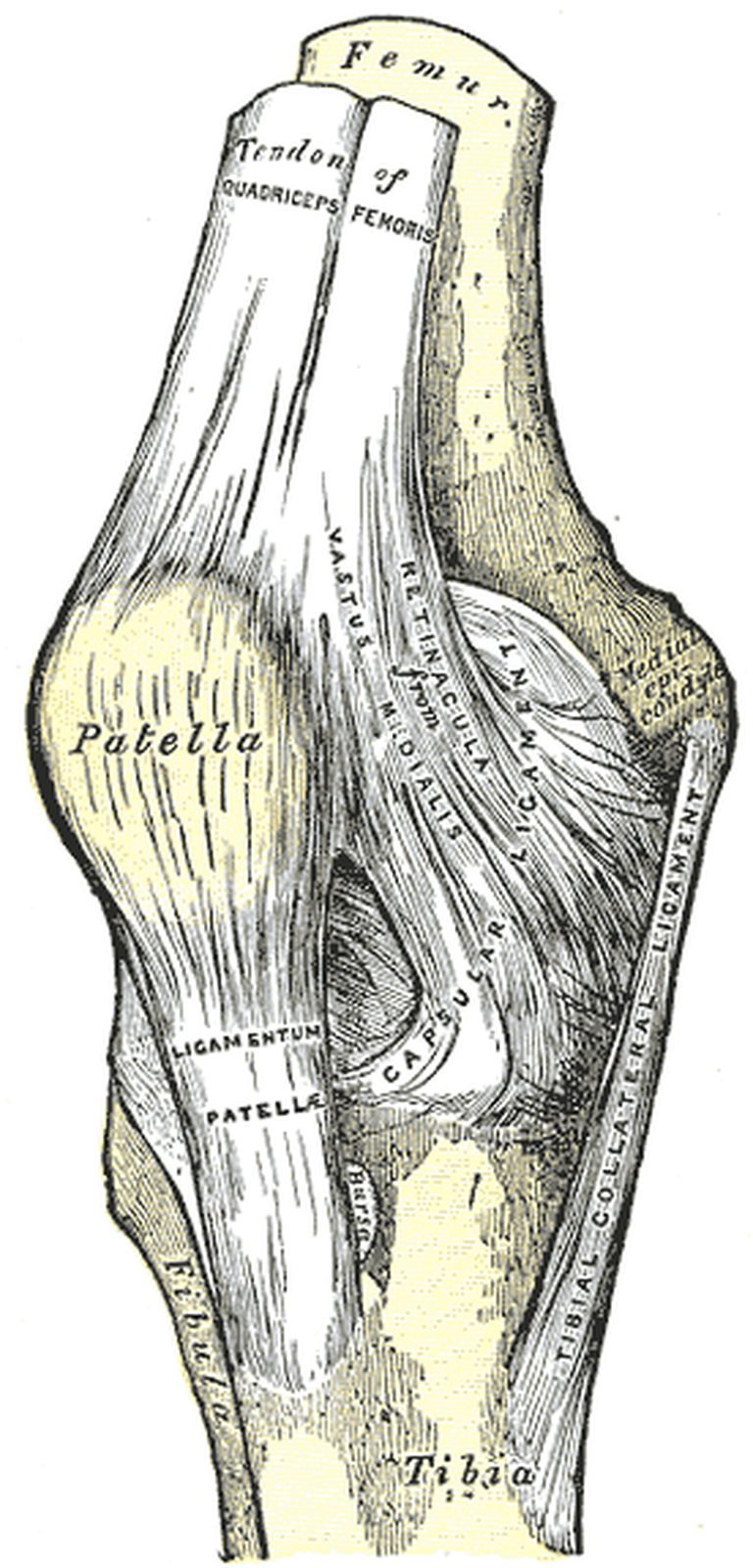
Knee joint from the front with the patellar ligament below the patella highlighted in yellow (Gray) Knee joint from the front with the patellar ligament below the patella highlighted in yellow (Gray)
Illustration: Henry Gray, Anatomy of the Human Body (1918), public domain · Highlighting: AnatomyProf

The patellar ligament continues the tendon of the quadriceps femoris muscle; the patella, the largest sesamoid bone of the body, is embedded in this tendon.

  • Course: from the apex of the patella to the tibial tuberosity; superficial fibres of the quadriceps tendon pass over the anterior surface of the patella into the ligament.
  • Function: reinforces the patella and thus the knee joint as a whole; transmits the force of the quadriceps to the tibia (extensor mechanism).

Behind the ligament lie the infrapatellar fat pad (Hoffa’s fat pad) and the deep infrapatellar bursa. Tapping the ligament elicits the patellar tendon reflex (femoral nerve, segments L3–L4). Its middle third is often used as a graft to reconstruct the anterior cruciate ligament. In the figure (knee from the front) the patellar ligament is highlighted in yellow.

Patellar retinacula

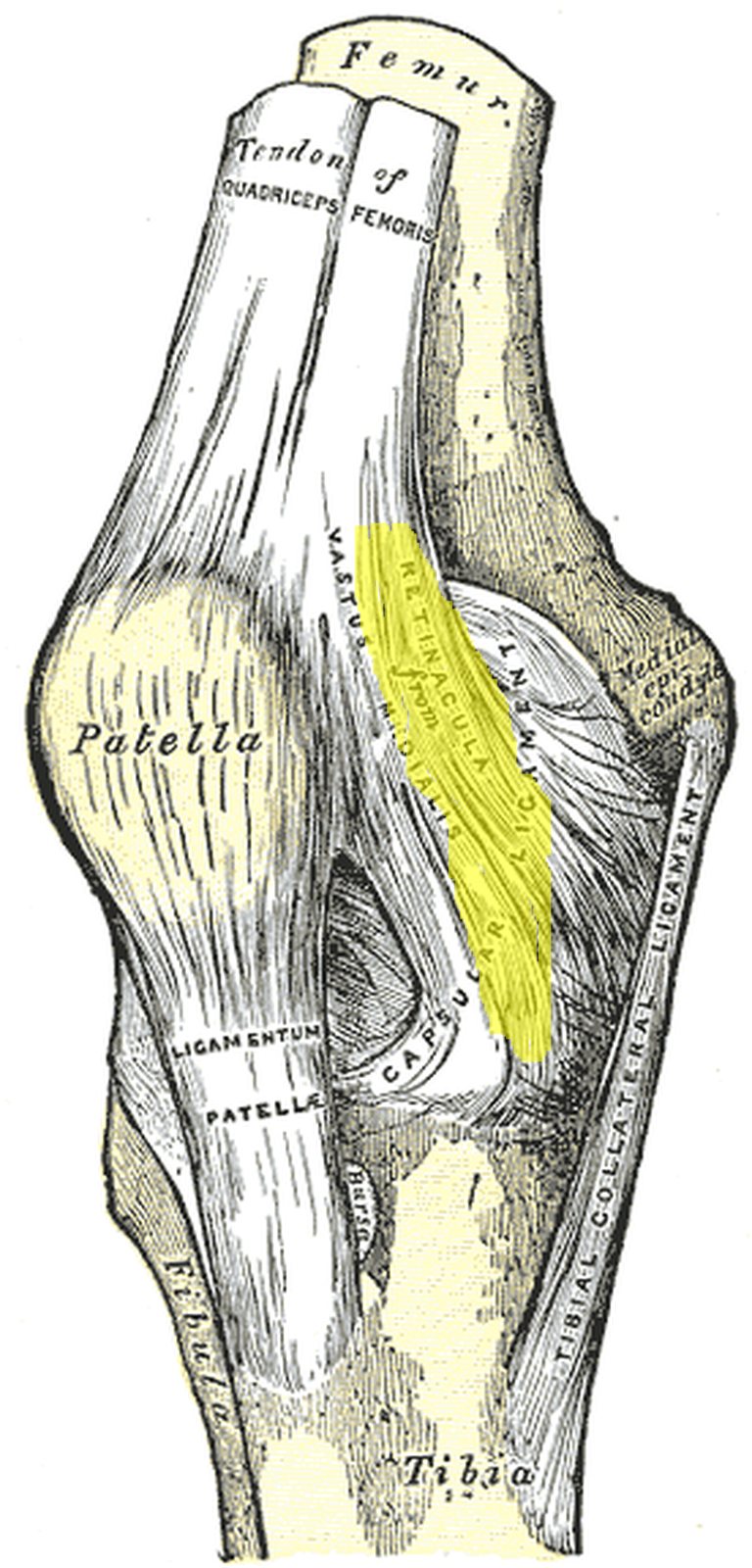
Knee joint from the front with the medial patellar retinaculum highlighted in yellow (Gray) Knee joint from the front with the medial patellar retinaculum highlighted in yellow (Gray)
Illustration: Henry Gray, Anatomy of the Human Body (1918), public domain · Highlighting: AnatomyProf

The medial and lateral patellar retinacula are tendinous bands that pass distally on either side of the patella. They derive mainly from the vastus medialis and vastus lateralis muscles; laterally, fibres of the iliotibial tract also contribute.

  • Function: lateral and medial stabilisation of the patella.
  • Insertion: on the tibial condyles on either side of the tibial tuberosity.

The retinacula form an “accessory extensor mechanism”: after a transverse fracture of the patella, limited extension of the knee remains possible as long as they are intact. In the common lateral dislocation of the patella the medial restraints (medial retinaculum, medial patellofemoral ligament) are typically torn. In the figure the medial retinaculum is highlighted in yellow; Gray labels it “Retinacula from vastus medialis”.

Collateral ligaments

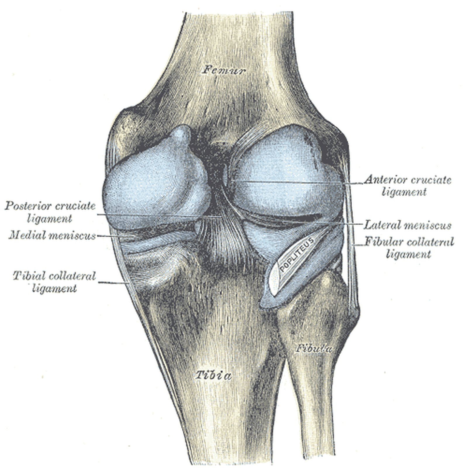
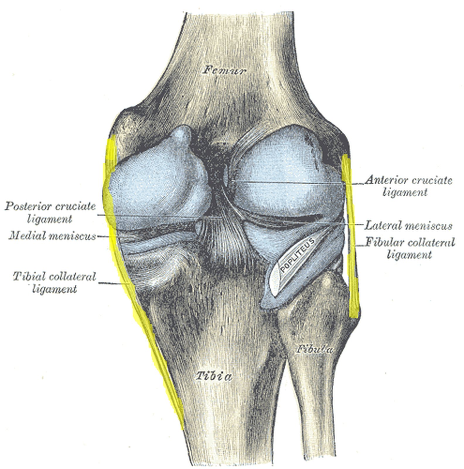
Knee joint from behind with the tibial and fibular collateral ligaments highlighted in yellow (Gray) Knee joint from behind with the tibial and fibular collateral ligaments highlighted in yellow (Gray)
Illustration: Henry Gray, Anatomy of the Human Body (1918), public domain · Highlighting: AnatomyProf

Tibial (medial) collateral ligament

  • Consists of two limbs, both arising from the medial epicondyle of the femur.
  • The anterior limb inserts on the medial surface of the tibia below the level of the tibial tuberosity; the posterior limb inserts posteriorly on the medial tibial condyle.
  • It is fused with the joint capsule and the medial meniscus; it prevents abduction (valgus) and limits rotation.

Fibular (lateral) collateral ligament

  • Lies outside the joint capsule and is not attached to the lateral meniscus.
  • Runs from the lateral epicondyle of the femur to the head of the fibula.
  • Prevents adduction (varus) and limits rotation.

Both collateral ligaments are taut in extension and slack in flexion – which is why rotation is only possible in the flexed knee; they mainly check external rotation. They are tested with valgus and varus stress. In the figure (posterior view) both collateral ligaments are highlighted in yellow.

Popliteal ligaments

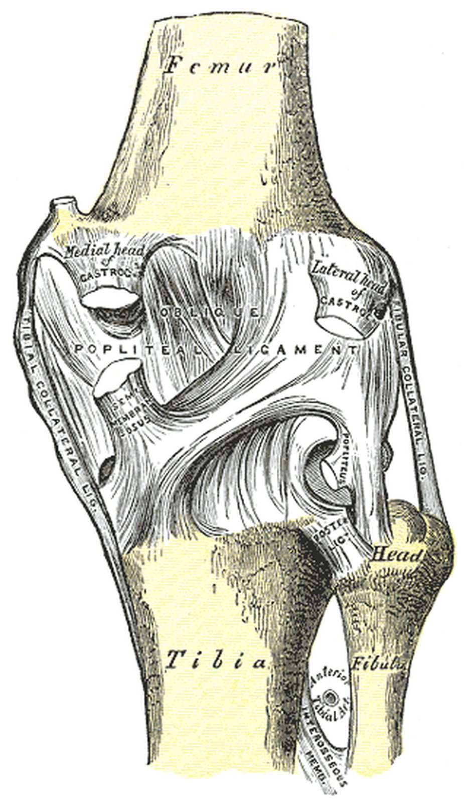
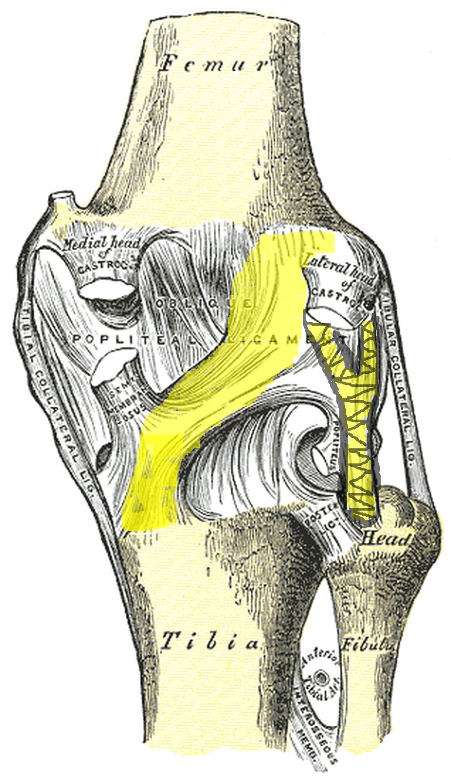
Knee joint from behind; oblique and arcuate popliteal ligaments highlighted in yellow (Gray) Knee joint from behind; oblique and arcuate popliteal ligaments highlighted in yellow (Gray)
Illustration: Henry Gray, Anatomy of the Human Body (1918), public domain · Highlighting: AnatomyProf

These ligaments lie in the popliteal fossa (back of the knee) and reinforce the posterior wall of the joint capsule. They prevent hyperextension; the oblique popliteal ligament also checks external rotation.

  • Oblique popliteal ligament: arises from the medial tibial condyle – it is an expansion of the semimembranosus tendon – and runs obliquely upwards to the lateral femoral condyle (lateral epicondyle).
  • Arcuate popliteal ligament: begins at the head of the fibula, arches over the popliteus tendon and radiates upwards and medially into the joint capsule.

Together with the semimembranosus tendon and the popliteus complex they form the posterior capsular restraint (“posterior corner”). A clinically important finding here is the Baker’s cyst, a fluid-filled outpouching of the joint between the semimembranosus and the medial head of the gastrocnemius. In the figure (posterior view) the obliquely running oblique popliteal ligament and the arcuate popliteal ligament rising from the fibular head are highlighted in yellow.

Test your knowledge

Multiple-choice questions on structure and function.

Updated: