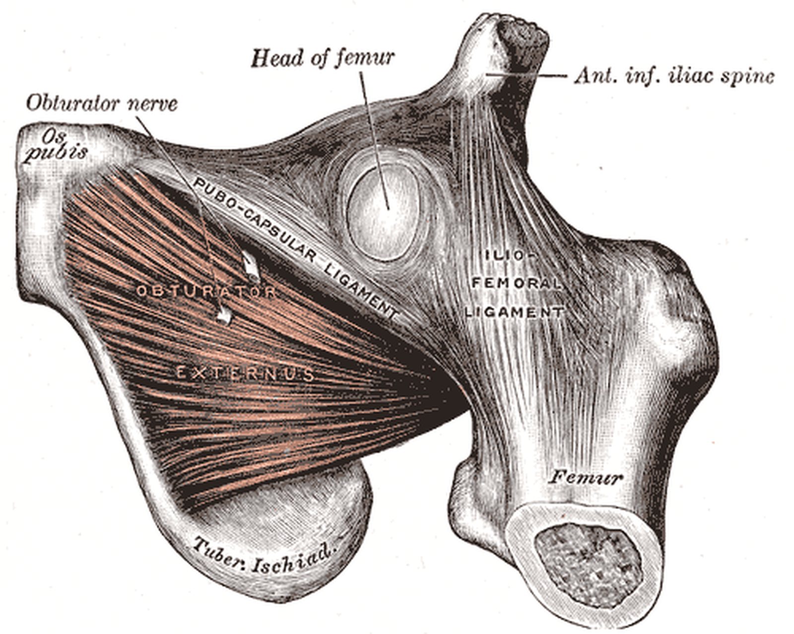
Ligaments of the hip joint

Hip joint

Start quiz

This unit covers the ligaments of the hip joint. They stabilise the joint and also limit movements of the hip joint (articulatio coxae). The iliofemoral ligament, ischiofemoral ligament, pubofemoral ligament and zona orbicularis are presented.

The hip is a ball-and-socket joint of the special “enarthrosis” type: the acetabulum embraces the femoral head beyond its equator. The three strong capsular ligaments wind spirally around the femoral neck. In extension they tighten, press the head into the socket and thus allow upright standing with little muscular effort.

Key facts

  • The iliofemoral ligament (anterior inferior iliac spine → intertrochanteric line) is the strongest ligament of the human body.
  • All three capsular ligaments run spirally and tighten in extension.
  • Iliofemoral ligament limits extension and adduction, pubofemoral mainly abduction, ischiofemoral internal rotation and extension.
  • The zona orbicularis encircles the femoral neck and keeps the femoral head in the socket.
  • Weak, ligament-free areas of the capsule lie between the ligaments, e.g. at the iliopectineal bursa.

Iliofemoral ligament
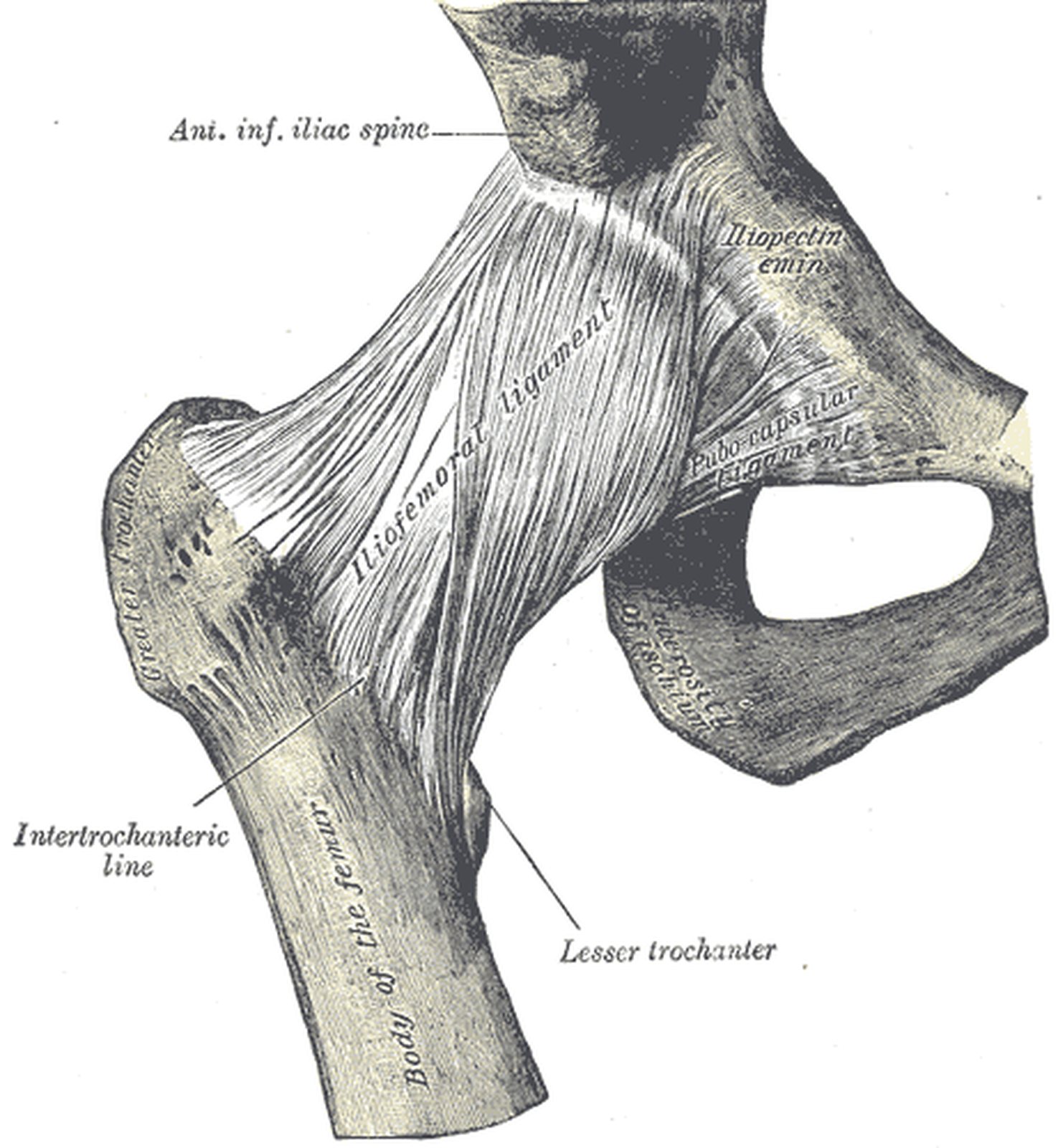
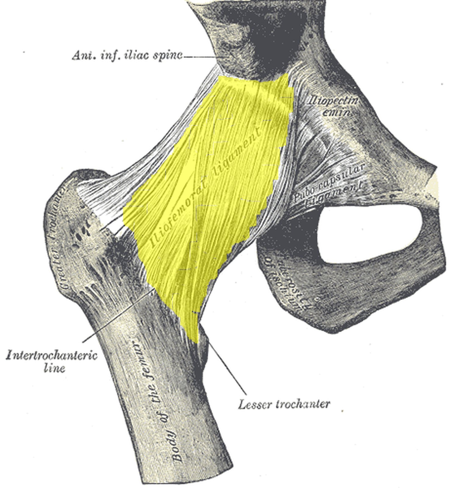
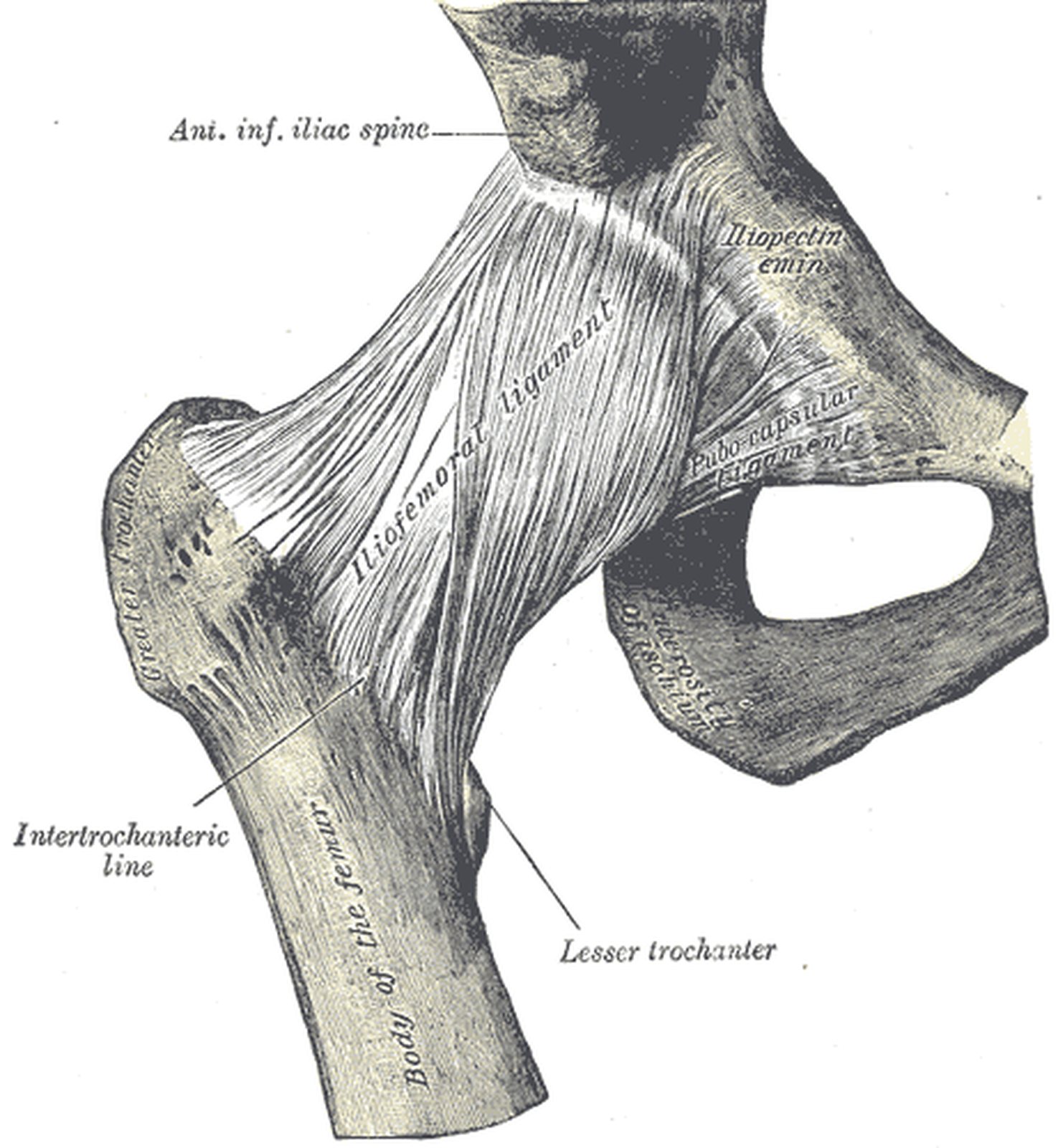
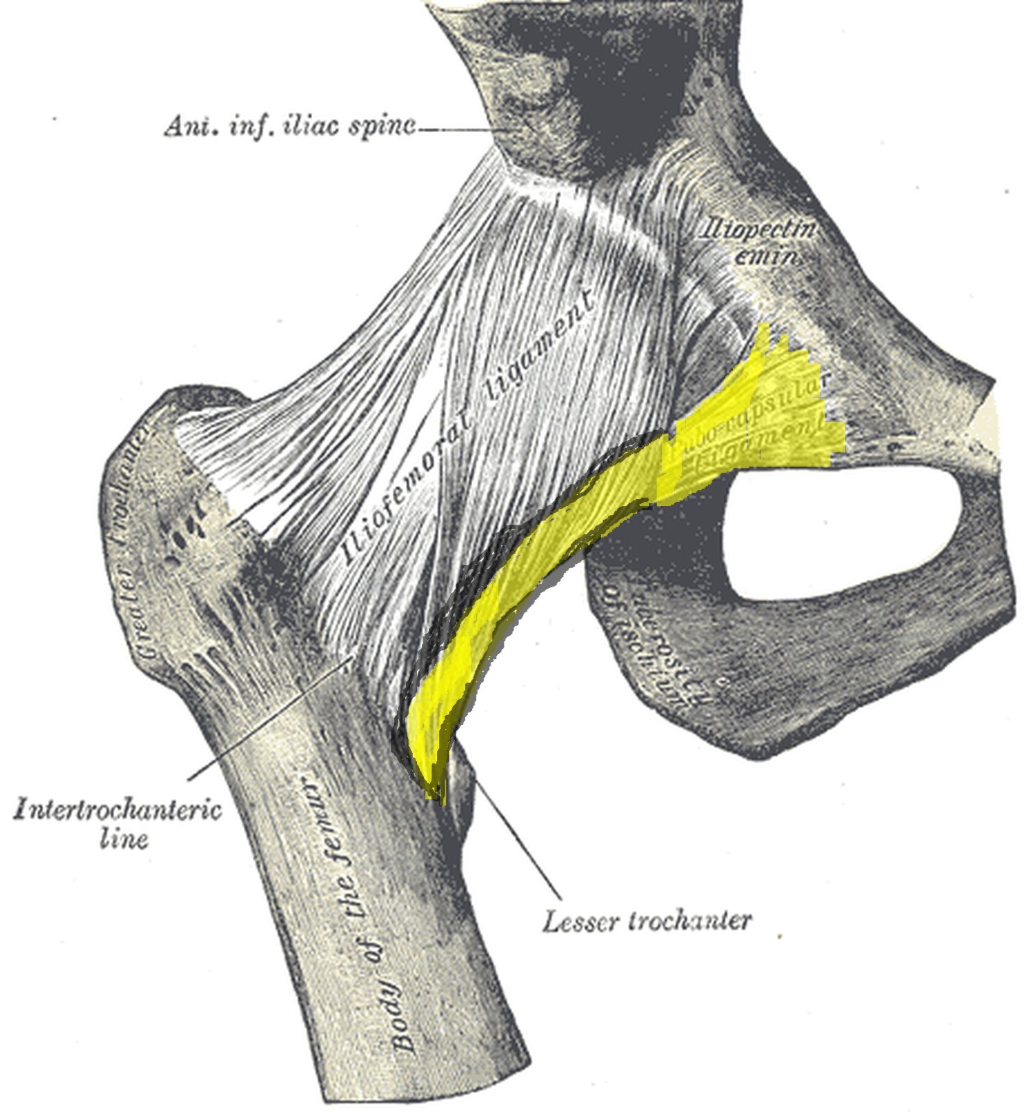
Right hip joint from the front with the iliofemoral ligament highlighted in yellow (Gray) Right hip joint from the front with the iliofemoral ligament highlighted in yellow (Gray)
Illustration: Henry Gray, Anatomy of the Human Body (1918), public domain · Highlighting: AnatomyProf

The iliofemoral ligament is the strongest ligament of the human body; it withstands tensile forces of several thousand newtons.

  • Course: from the anterior inferior iliac spine (ilium) to the intertrochanteric line.
  • Function: limits extension and adduction.

The ligament has the shape of an inverted Y (Y-ligament of Bigelow) with a lateral and a medial part. The lateral part mainly limits adduction and external rotation, the medial part extension. In the upright stance it prevents the pelvis from tilting backwards – the body effectively “hangs” on its iliofemoral ligaments. In posterior dislocation of the hip the ligament usually remains intact and determines the typical position of the limb (flexion, adduction, internal rotation). In the figure (hip joint from the front) the iliofemoral ligament is highlighted in yellow.

Ischiofemoral ligament

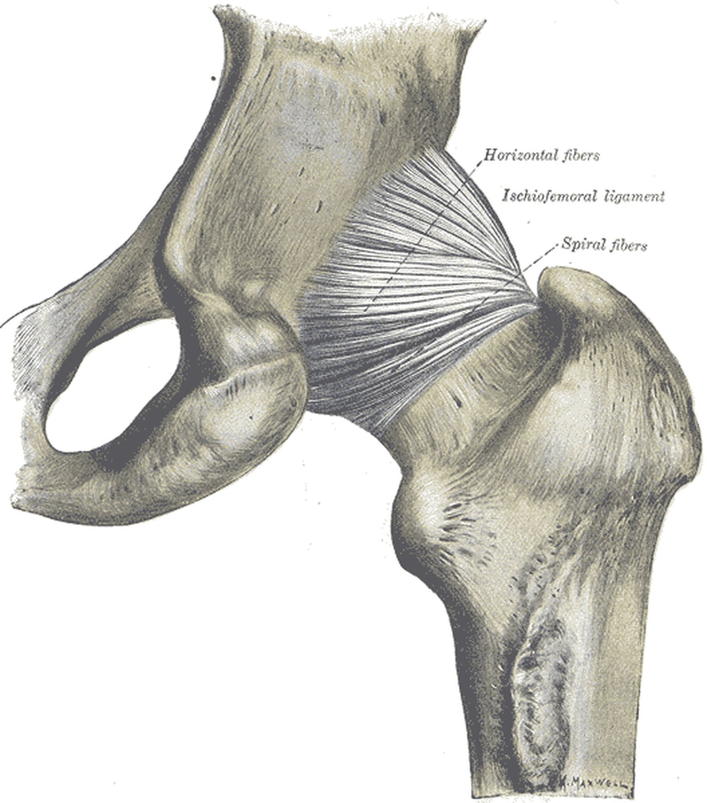
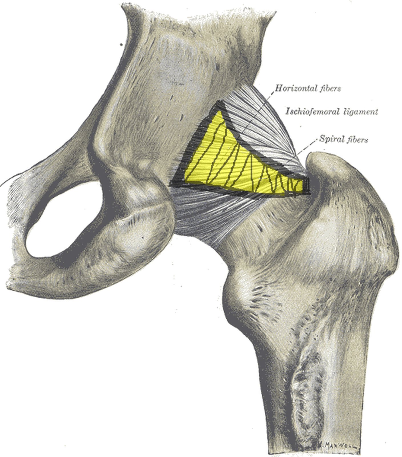
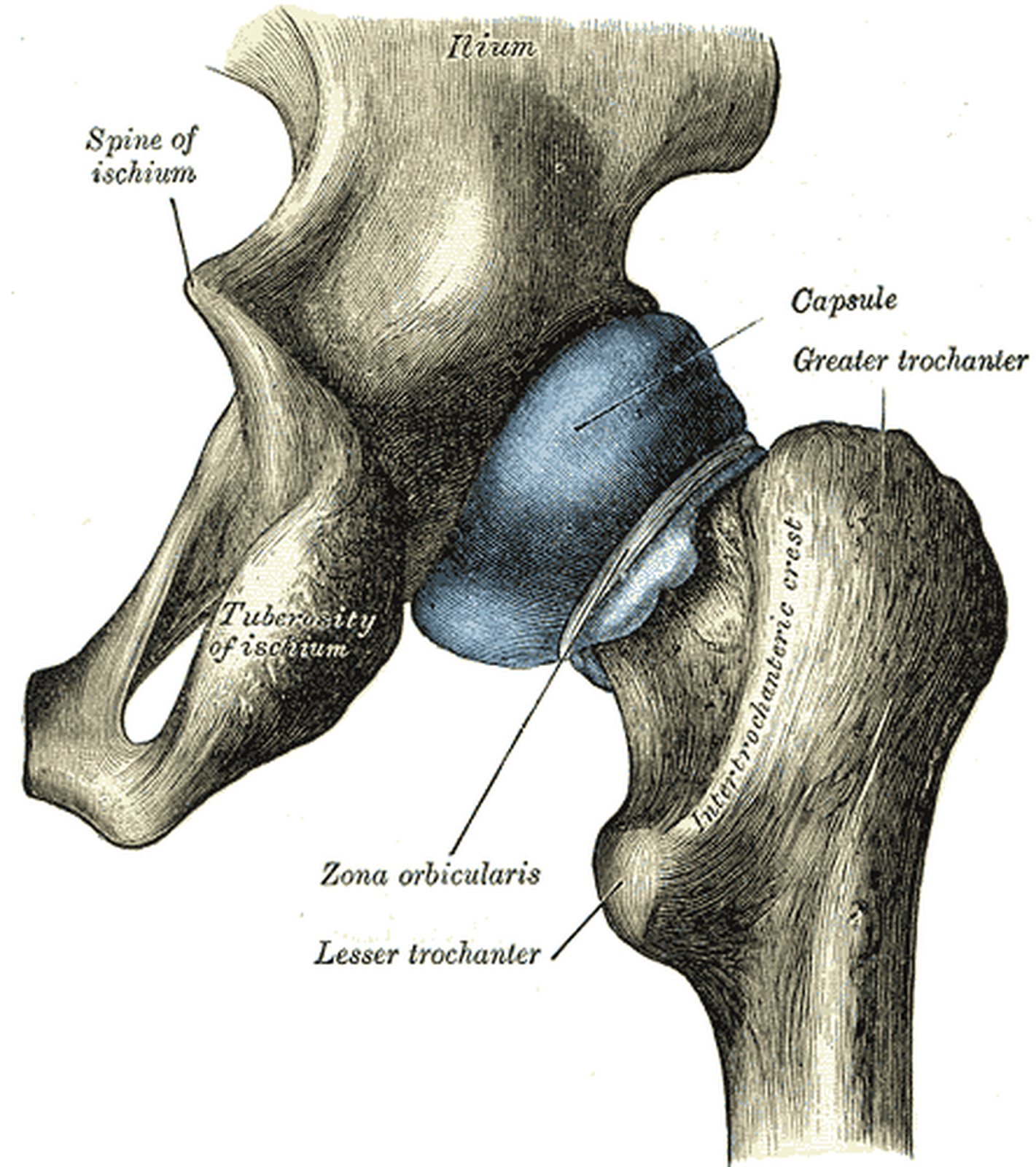
Hip joint from behind with the ischiofemoral ligament highlighted in yellow (Gray) Hip joint from behind with the ischiofemoral ligament highlighted in yellow (Gray)
Illustration: Henry Gray, Anatomy of the Human Body (1918), public domain · Highlighting: AnatomyProf

The ischiofemoral ligament is the weakest of the three capsular ligaments and lies on the posterior aspect of the joint.

  • Course: from the ischium (posterior rim of the acetabulum) to the intertrochanteric crest / trochanteric fossa on the posterior aspect of the femur; some fibres blend with the zona orbicularis.
  • Function: limits internal rotation and extension, and adduction when the hip is flexed.

Its fibres spiral from posteroinferior to superolateral around the femoral neck. In extension they are “wound up” and tightened like the other capsular ligaments. In the figure (hip joint from behind) the ischiofemoral ligament is highlighted in yellow; Gray distinguishes its “horizontal fibers” and “spiral fibers”.

Pubofemoral ligament

Hip joint from the front with the pubofemoral ligament (Gray: pubocapsular ligament) highlighted in yellow Hip joint from the front with the pubofemoral ligament (Gray: pubocapsular ligament) highlighted in yellow
Illustration: Henry Gray, Anatomy of the Human Body (1918), public domain · Highlighting: AnatomyProf

The pubofemoral ligament arises from the pubis and reinforces the capsule anteroinferiorly.

  • Course: from the superior pubic ramus to the distal part of the intertrochanteric line of the femur; its fibres blend with the capsule and the medial part of the iliofemoral ligament.
  • Function: mainly limits abduction of the femur, and also extension and external rotation.

Between the pubofemoral and iliofemoral ligaments lies a gap where the iliopectineal (iliopsoas) bursa often communicates with the joint cavity. A second weak point lies posteroinferiorly between the pubofemoral and ischiofemoral ligaments. In the figure (hip joint from the front) the pubofemoral ligament is highlighted in yellow; Gray uses the older name “Pubocapsular ligament”.

Zona orbicularis

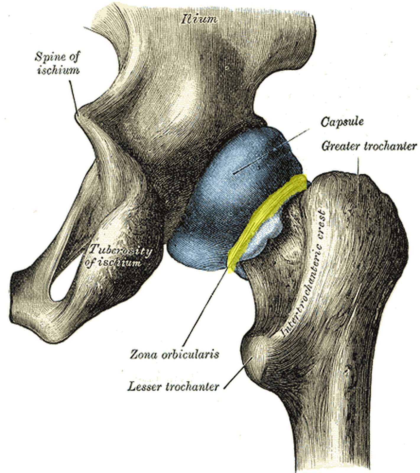
Hip joint from behind with blue capsule and the zona orbicularis around the femoral neck in yellow (Gray) Hip joint from behind with blue capsule and the zona orbicularis around the femoral neck in yellow (Gray)
Illustration: Henry Gray, Anatomy of the Human Body (1918), public domain · Highlighting: AnatomyProf

The zona orbicularis (annular zone) is a ring of fibres in the deep layers of the joint capsule.

  • It is formed circularly by fibres of the iliofemoral, ischiofemoral and pubofemoral ligaments.
  • It lies distal to the femoral head and encircles the femoral neck at its narrowest point.
  • This annular zone prevents the femoral head from slipping out of the socket (acetabulum).

Because the ring is narrower than the diameter of the femoral head, it holds the head like a buttonhole. The zona orbicularis has no bony attachment; it only becomes clearly visible once the capsular ligaments have been removed. The retinacular arteries run to the femoral head within the capsule along the neck – which is why femoral neck fractures carry a risk of avascular necrosis of the head. In the figure (hip joint from behind, capsule in blue) the zona orbicularis is highlighted in yellow.

Test your knowledge

Multiple-choice questions on structure and function.

Updated: