Dense regular connective tissue
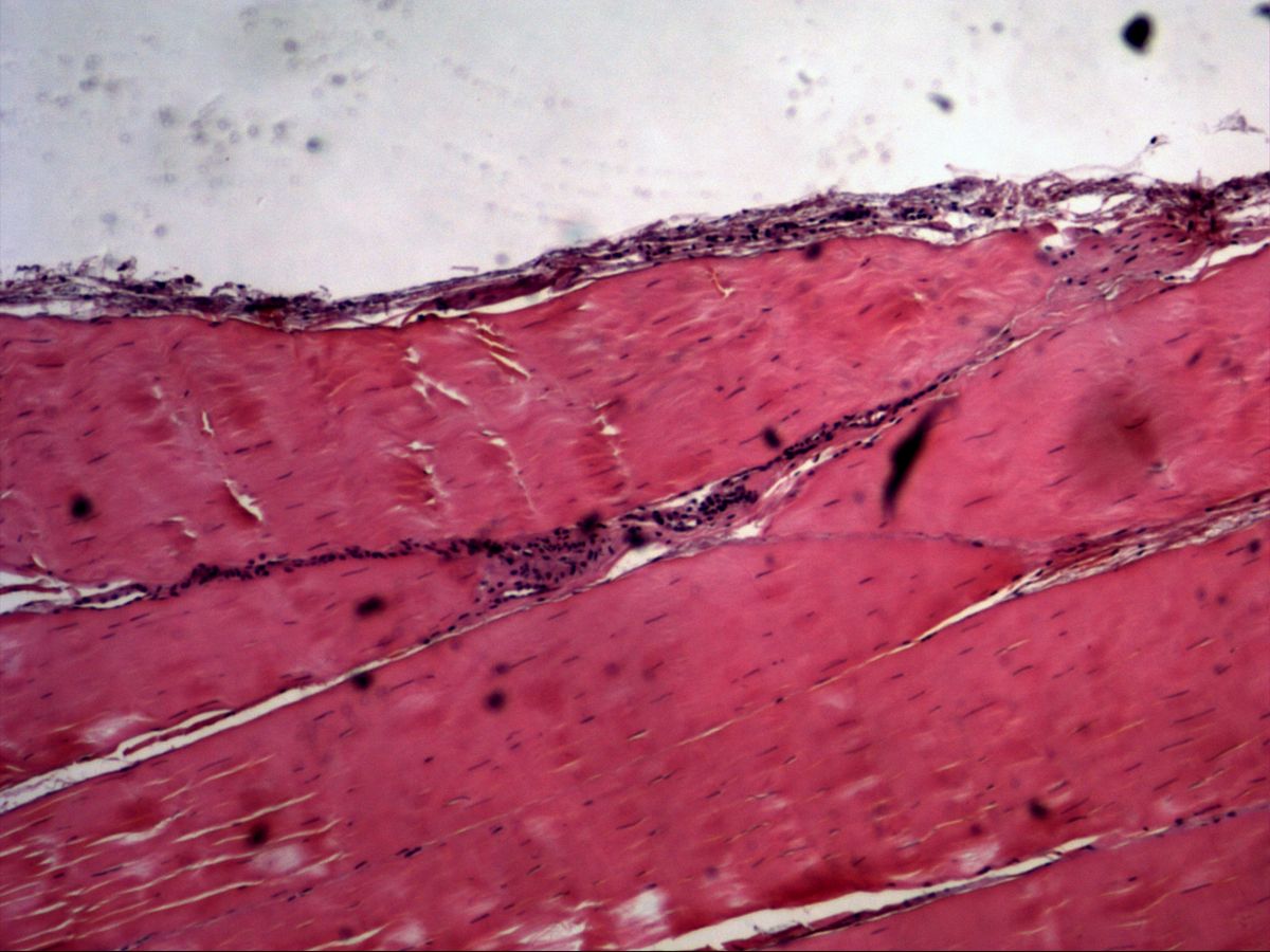
Tendon, longitudinal · H&E
Labelled structures
This image has no labels – use the caption instead.
Dense regular connective tissue is optimised for tension in a single direction. Its typical example is the tendon: densely packed, parallel type I collagen bundles transmit muscle force to bone. Flattened tendon cells lie in rows between the bundles.
How to recognise it
- Densely packed, parallel, strongly eosinophilic collagen bundles
- Long, slender, dark nuclei of wing cells in rows between the bundles
- Little ground substance, few vessels
- Pale clefts or loose connective tissue between bundles (internal peritendineum)
- More cellular sheath at the edge (external peritendineum)
Structures to find
- Parallel collagen bundles
- Wing cells (tenocytes) and their nuclei
- Internal peritendineum
- External peritendineum
- Small vessels in the peritendineum
Structure
Collagen fibrils assemble into fibres and these into primary bundles. Each primary bundle is surrounded by delicate loose connective tissue, the internal peritendineum (endotenon); several primary bundles form secondary bundles. The whole tendon is enclosed by the denser external peritendineum (epitenon), through which vessels and nerves enter.
Tendon cells (tenocytes) are specialised fibrocytes. Squeezed between the fibre bundles, they form thin, wing-like cytoplasmic sheets – hence the name wing cells. In longitudinal section only their long, slender nuclei in rows are visible.
Function and occurrence
The parallel alignment of the collagen bundles makes tendons extremely resistant to tension but hardly extensible. The slightly wavy fibre pattern (crimp) is straightened first under low load, buffering peaks of tension. Dense regular connective tissue forms tendons, aponeuroses and most ligaments. A special form are elastic ligaments such as the ligamenta flava, which consist mainly of elastic fibres. Tendons are poorly vascularised and therefore have a slow metabolism.
Microscopy tips
In longitudinal section the picture is very uniform: strongly eosinophilic parallel fibre bundles with slender, dark, rod-shaped nuclei in rows between them. The pale longitudinal clefts between the bundles correspond to the internal peritendineum or shrinkage artefacts. At the edge you will find the more cellular external peritendineum. Pitfall – smooth muscle: its nuclei lie in the centre of the cell, are cigar-shaped with rounded ends, and the cytoplasm is less bright pink. Skeletal muscle shows cross-striations and peripheral nuclei.
Clinical relevance
Because of the poor blood supply, tendon injuries heal slowly; the repair tissue is initially disorganised and less resilient. Chronic overload causes tendinopathy with degeneration of the fibre bundles, e.g. of the Achilles or supraspinatus tendon. A degeneratively damaged Achilles tendon may rupture under sudden load. Fluoroquinolone antibiotics and glucocorticoids also increase the risk of rupture. Tendon reconstruction often uses autologous grafts, e.g. from the semitendinosus or patellar tendon in cruciate ligament replacement; the graft is slowly repopulated and remodelled by host cells.
Slide images
Test your knowledge
Assign the correct number in the image to each term.
Tap the requested structure in the image.
Multiple-choice questions on structure and function.
Frequently asked questions
Why are tendon cells called wing cells?
They lie between the collagen bundles and extend thin, plate-like cytoplasmic processes between the bundles. In cross-section they therefore look stellate with wing-like extensions.
How do I tell tendon from smooth muscle?
In tendon the nuclei are very slender and dark and lie between brightly eosinophilic fibres. Smooth muscle cells have central, cigar-shaped nuclei with rounded ends and paler cytoplasm.
What is the peritendineum?
The internal peritendineum is loose connective tissue around the fibre bundles, carrying vessels and nerves. The external peritendineum envelops the whole tendon.
Related topics
Updated: