Loose and dense irregular connective tissue
Skin · H&E
Labelled structures
This image has no labels – use the caption instead.
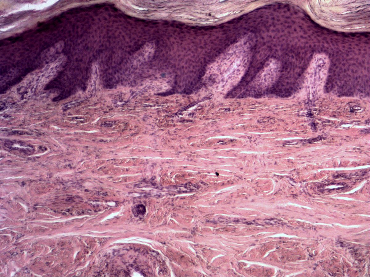
The dermis of the skin shows two types of connective tissue stacked on top of each other: beneath the epidermis lies the cell- and vessel-rich loose connective tissue of the papillary layer, below it the fibre-rich dense irregular connective tissue of the reticular layer. Skin is therefore ideal for comparing both forms in a single section.
How to recognise it
- Two layers: pale, cell-rich papillary layer above strongly pink reticular layer
- Loose CT: fine, loosely spaced fibres, abundant ground substance, many capillaries
- Loose CT: numerous free cells with round nuclei
- Dense irregular CT: thick collagen bundles in all directions, cut transversely, obliquely and longitudinally
- Few slender, dark fibrocyte nuclei between the bundles
Structures to find
- Epidermis and basement membrane (orientation)
- Dermal papillae with loose connective tissue
- Fibroblasts and fibrocytes
- Free cells (e.g. lymphocytes)
- Loose collagen fibres (types I and III)
- Interwoven collagen bundles in the reticular layer
- Capillaries and small vessels
Structure
Connective tissue consists of cells and extracellular matrix (fibres and ground substance). In loose connective tissue ground substance and cells predominate: resident fibroblasts or fibrocytes plus numerous free cells such as lymphocytes, macrophages, mast cells and plasma cells. The collagen fibres (mainly type I, also type III) are thin and loosely arranged, with capillaries in between.
In dense irregular connective tissue thick collagen bundles dominate and interweave in all directions. Cells and ground substance recede; the few fibrocyte nuclei lie slender between the bundles.
Function and occurrence
Loose connective tissue is the body's packing and defence tissue: it fills spaces, accompanies vessels and nerves, binds water and is the site of immune reactions and inflammation. Typical locations are the papillary dermis, the lamina propria of mucous membranes and the interstitium of organs.
Dense irregular connective tissue resists tensile forces acting from changing directions. It forms the reticular dermis, organ capsules, periosteum, perichondrium, dura mater and sclera.
Microscopy tips
Start at the epidermis at low power and move downwards: immediately beneath the basement membrane and in the dermal papillae the tissue looks paler, cell-rich and finely fibrous – this is loose connective tissue. Below, the dermis suddenly turns strongly pink with thick, wavy interwoven bundles – dense irregular connective tissue.
- Fibrocyte nuclei: slender, dark, spindle-shaped
- Free cells: round nuclei, especially around vessels
- White clefts between bundles are mostly shrinkage artefacts
Differential: tendon has parallel bundles; smooth muscle has cigar-shaped nuclei in the centre of the cells.
Clinical relevance
Because loose connective tissue binds a lot of water, fluid accumulates here in oedema, and inflammation spreads rapidly through it (e.g. cellulitis). In disorders of collagen synthesis such as Ehlers–Danlos syndrome the tensile strength of dense connective tissue in particular is reduced: the skin is hyperextensible and fragile, and wounds heal with wide scars. Scar tissue itself is dense connective tissue, initially rich in type III collagen.
Slide images
Test your knowledge
Assign the correct number in the image to each term.
Tap the requested structure in the image.
Multiple-choice questions on structure and function.
Frequently asked questions
What is the difference between a fibroblast and a fibrocyte?
Fibroblasts are the active form that synthesises fibres and matrix, with a larger, paler nucleus and basophilic cytoplasm. Fibrocytes are the resting form with a slender, dark nucleus and barely visible cytoplasm; the two forms can convert into each other.
Why is the papillary layer loose and the reticular layer dense?
The papillary layer nourishes the avascular epidermis and serves defence, so it needs capillaries, ground substance and free cells. The reticular layer gives the skin its tensile strength against pull from all directions.
How do I tell dense irregular from dense regular connective tissue?
In irregular tissue the bundles in one section are cut transversely, obliquely and longitudinally. In regular tissue (tendon) all bundles and fibrocyte nuclei run in the same direction.
Related topics
Updated: