Keratinised stratified squamous epithelium
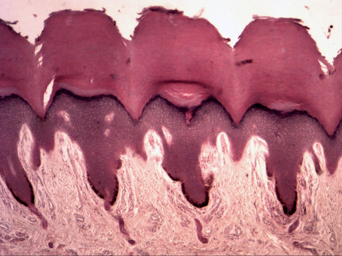
Fingertip skin · H&E
Labelled structures
This image has no labels – use the caption instead.
In humans, keratinised stratified squamous epithelium is found exclusively in the epidermis. The skin is called the cutis, the underlying tissue the subcutis (fat and loose connective tissue, mechanoreceptors, nerves). The cutis is divided into the epidermis and the dermis/corium. The epithelium (epidermis) consists of the layers stratum corneum, stratum lucidum, stratum granulosum, stratum spinosum and stratum basale. Connective tissue fibres radiating into the basement membrane are attached by anchoring fibrils. The dermis contains the stratum papillare as well as the sebaceous and scent glands.
The fingertip belongs to thick (ridged) skin: it is hairless, has no sebaceous glands, but numerous eccrine sweat glands and an exceptionally thick horny layer. Epidermal ridges reach deep into the dermis, with tall connective tissue papillae in between. This makes it easy to follow every layer of the epidermis here.
How to recognise it
- Very thick, anuclear, homogeneously pink horny layer
- Dark line of the stratum granulosum
- Polygonal cells in the stratum spinosum
- Deep epidermal ridges and tall dermal papillae
- No hairs and no sebaceous glands
Structures to find
- Stratum corneum
- Stratum granulosum
- Stratum spinosum
- Stratum basale
- Basement membrane
- Papillary layer of the dermis
Structure
From basal to apical: the stratum basale is one layer of columnar cells with dark nuclei on the basement membrane (stem cells, with melanocytes in between). The stratum spinosum contains several layers of polygonal cells joined by desmosomes. The stratum granulosum consists of a few layers of flattened cells with basophilic keratohyalin granules – a dark line in the slide. In thick skin a narrow, homogeneous stratum lucidum may lie above it. The stratum corneum consists of anuclear, keratin-filled horny cells.
Function
Keratinocytes migrate from the base to the surface in about four weeks, losing their nucleus and organelles and becoming horny cells (keratinisation). Lipids from the lamellar bodies of the stratum granulosum seal the intercellular spaces. This creates a barrier against water loss, microbes and mechanical stress. The interlocking of epidermal ridges and dermal papillae anchors the epidermis and provides space for Meissner corpuscles – the basis of the fine sense of touch in the fingertip.
Microscopy tips
At low magnification the broad, homogeneously pink horny layer stands out, often thicker than the living part of the epidermis. The dark line beneath it is the stratum granulosum – a good landmark. At high magnification you see round nuclei with a pale halo in the stratum spinosum and the basal cells lining the papillae. Corkscrew-like channels in the horny layer are sweat gland ducts. Differential diagnosis from the oesophagus: there the horny layer and stratum granulosum are absent and surface cells have nuclei.
Clinical relevance
The ridge patterns of the fingertip are genetically determined and unchanged throughout life – the basis of fingerprints. In psoriasis cell turnover is greatly accelerated: the horny layer still contains nuclei (parakeratosis) and the stratum granulosum is missing. Basal cell and squamous cell carcinomas can arise from basal cells and keratinocytes. Calluses (hyperkeratosis) show how the horny layer responds to chronic pressure by thickening.
Slide images
Test your knowledge
Assign the correct number in the image to each term.
Tap the requested structure in the image.
Multiple-choice questions on structure and function.
Frequently asked questions
Why is the stratum granulosum so dark?
Its cells contain basophilic keratohyalin granules that stain strongly with haematoxylin. The layer therefore appears as a dark band beneath the horny layer.
Are there sebaceous glands in fingertip skin?
No. Sebaceous glands are associated with hair follicles, and the ridged skin of the hand is hairless. Instead it has particularly many eccrine sweat glands.
Is the stratum lucidum always visible?
No, it occurs only in thick skin and is often not distinguishable in H&E sections. If it is absent in the slide, that is not an error.
Related topics
Updated: