Simple squamous and simple cuboidal epithelium
Kidney · H&E
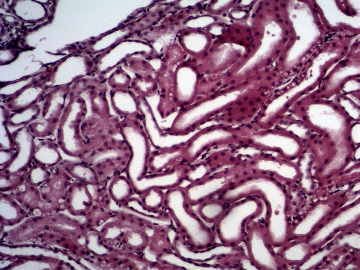
Labelled structures
This image has no labels – use the caption instead.
A histological section of kidney tissue. Tubules can be seen. Collecting ducts cannot be identified here – they are larger and their cell borders are easier to distinguish.
The kidney is the classic slide for seeing the two simplest covering epithelia side by side: simple cuboidal epithelium with cube-shaped cells and round, central nuclei in many tubule segments, and simple squamous epithelium with flat, spindle-shaped nuclei in the thin limbs of the loop of Henle and in capillary endothelium.
How to recognise it
- Only one cell layer, all cells on the basement membrane
- Cuboidal: cells about as tall as wide, round central nucleus
- Squamous: very flat cells with spindle-shaped nucleus
- Tubules seen as rings of epithelial cells around a lumen
- Capillaries with erythrocytes between the tubules
Structures to find
- Tubule with simple cuboidal epithelium
- Tubule lumen
- Nuclei with hetero- and euchromatin
- Cell borders
- Basement membrane
- Capillary with endothelial nucleus and erythrocytes
Structure
Simple epithelia consist of a single cell layer that rests entirely on the basement membrane. In simple cuboidal epithelium the cells are roughly as tall as they are wide; the nuclei are round, central and show dark heterochromatin next to pale euchromatin. In simple squamous epithelium the cells are very flat, and the flattened nucleus often bulges into the lumen. In cross-section a tubule appears as a ring of epithelial cells around a central lumen, surrounded by basement membrane and interstitial connective tissue with capillaries.
Function and occurrence
Flat cells provide a minimal diffusion distance, so squamous epithelium is found wherever substances or gases are exchanged rapidly – endothelium of blood vessels, mesothelium of serous membranes, alveolar epithelium and the thin limbs of the loop of Henle. Cuboidal epithelium has more cytoplasm and room for organelles; it performs transport and secretion, e.g. in renal tubules and collecting ducts, small gland ducts, thyroid follicles and on the surface of the ovary.
Microscopy tips
At medium magnification find an area with tubules cut in cross-section, then switch to high power. Count the cell layers: if every nucleus touches the basement membrane, the epithelium is simple. Watch the nuclear shape – round and central suggests cuboidal, flat and spindle-shaped suggests squamous. You can tell capillaries from thin tubules by erythrocytes in the lumen and usually only one or two endothelial nuclei per profile. Proximal tubules look 'fuzzy' towards the lumen because of their brush border and strongly eosinophilic cytoplasm, whereas collecting ducts show clearly visible cell borders.
Clinical relevance
Tubular epithelia are highly sensitive to hypoxia and nephrotoxic substances. In acute tubular necrosis (e.g. after shock or due to drugs) epithelial cells detach, nuclei are lost and the lumen fills with cell debris, so the intact single row of cells is disrupted. Because the epithelium can regenerate, this damage is often reversible. The squamous endothelium of capillaries is clinically relevant too: damage to glomerular endothelium leads to loss of protein and blood into the urine.
Slide images
Test your knowledge
Assign the correct number in the image to each term.
Tap the requested structure in the image.
Multiple-choice questions on structure and function.
Frequently asked questions
How do I distinguish simple cuboidal from simple squamous epithelium?
By the ratio of cell height to width and by nuclear shape: cuboidal cells are cube-shaped with a round nucleus, squamous cells are flat with a flattened, spindle-shaped nucleus.
Why does the lumen of proximal tubules often look 'fuzzy'?
The cells carry a dense brush border of microvilli, which appears in the light microscope as a fine eosinophilic fringe that partly fills the lumen.
Is endothelium an epithelium?
Yes, functionally and morphologically endothelium is a simple squamous epithelium lining the vessels. It is often named separately only because of its mesodermal origin.
Related topics
Updated: