Urothelium (transitional epithelium)
Urinary bladder · H&E
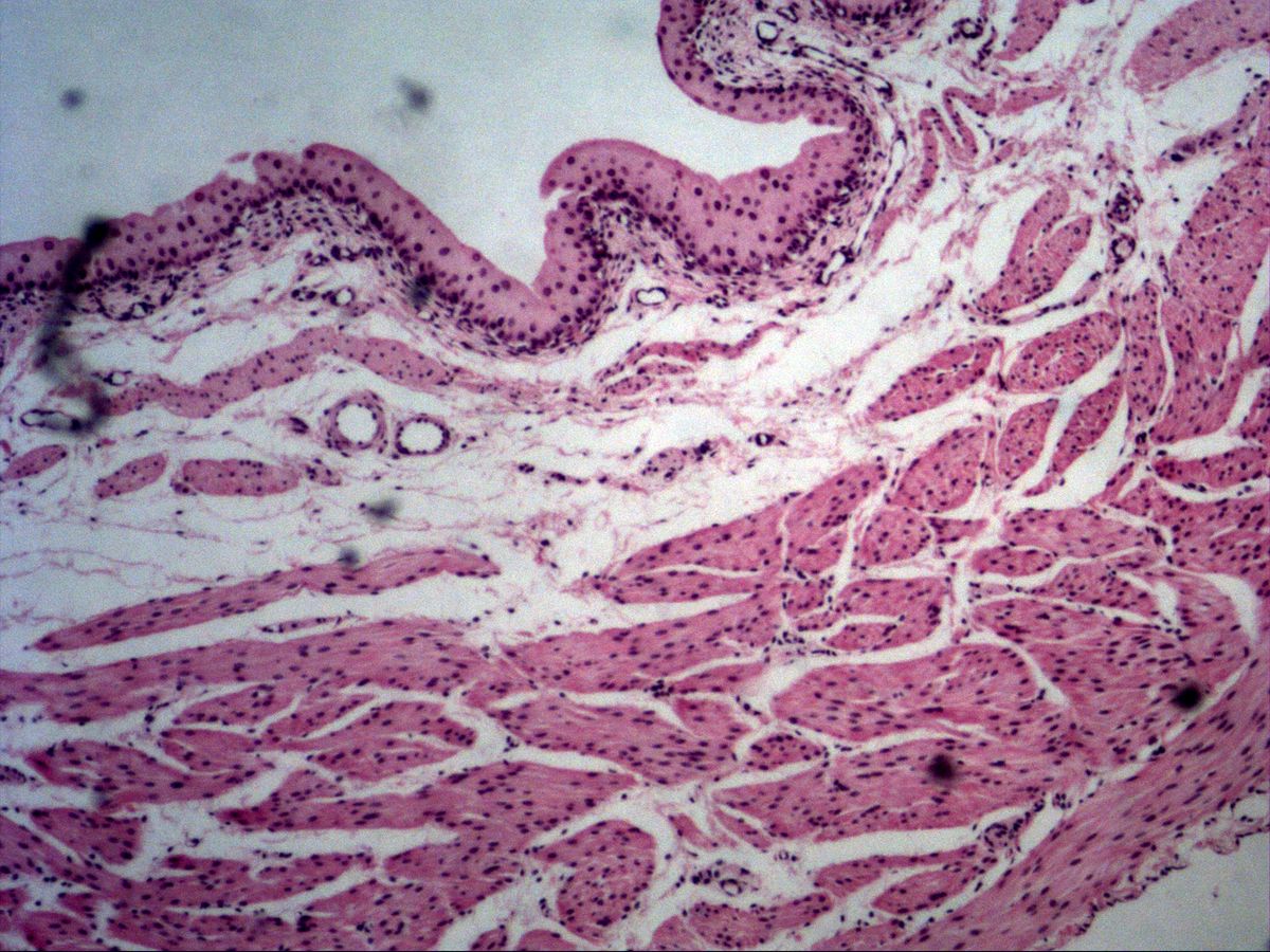
Labelled structures
This image has no labels – use the caption instead.
Transitional epithelium is found in the urinary tract. It can adapt to the filling level and has a protective crusta. Uroplakins and discoid vesicles strengthen this protection.
The urothelium is a specialised epithelium of the urinary tract (renal pelvis, ureter, bladder, proximal urethra). It consists of basal cells, several layers of intermediate cells and large umbrella cells, which often have two nuclei and cover several underlying cells. Depending on the degree of filling it looks tall and many-layered (empty bladder) or flat with only a few layers (full bladder).
How to recognise it
- Large dome-shaped umbrella cells at the surface
- Eosinophilic crusta on the luminal side
- Umbrella cells often binucleate
- Epithelial thickness depends on filling state
- Strong smooth muscle beneath, no muscularis mucosae
Structures to find
- Crusta
- Umbrella cells
- Intermediate cells
- Basal cells
- Basement membrane
- Lamina propria with blood vessels
- Muscular layer
Structure
The basal cells are small and rest on the basement membrane. Above them lie pear- to club-shaped intermediate cells, which according to current views reach the basement membrane with processes – the urothelium is therefore often regarded as pseudostratified. The superficial umbrella cells are very large, frequently binucleate or polyploid, and carry the crusta on their luminal side: a condensed, eosinophilic zone of cytoplasm with plaques of uroplakins in the apical membrane. Beneath the epithelium lie a vascular lamina propria and the strong smooth muscle (detrusor muscle).
Function
The urothelium forms one of the tightest barriers in the body: uroplakin plaques and tight junctions prevent water and urinary solutes from passing from the hypertonic urine into the tissue. When the bladder fills, discoid vesicles carrying prefabricated membrane are inserted into the apical surface, the cells flatten and the epithelium stretches without tearing. When the bladder empties, the membrane is endocytosed again. At the same time the intermediate cells slide past each other, so the number of visible cell layers decreases. In this way the urothelium protects the underlying tissue even when strongly stretched.
Microscopy tips
At low magnification the empty bladder shows a folded mucosa with a relatively thick epithelium over a loose lamina propria, followed by thick interwoven smooth muscle bundles. At high magnification look for the large dome-shaped umbrella cells with an eosinophilic rim (crusta) and occasionally two nuclei. Differential diagnosis from non-keratinised squamous epithelium: there the superficial cells become flat and small, whereas in urothelium they remain large and domed. A muscularis mucosae is absent.
Clinical relevance
Urothelial carcinoma is the most common tumour of the bladder; the most important risk factor is smoking, along with aromatic amines (occupational disease). The leading symptom is painless haematuria. In urinary tract infections bacteria attach via fimbriae to the uroplakins of umbrella cells. In a slide of a full bladder the urothelium may be only two or three cell layers thick and easily confused with squamous epithelium – then look for the crusta. Chronic irritation (e.g. bladder stones, schistosomiasis) can cause squamous metaplasia.
Test your knowledge
Assign the correct number in the image to each term.
Tap the requested structure in the image.
Multiple-choice questions on structure and function.
Frequently asked questions
Why is urothelium also called transitional epithelium?
It was once thought to be morphologically intermediate between stratified squamous and columnar epithelium. In fact it is a distinct epithelium that adapts its height to the filling state.
What is the crusta?
A condensed apical cytoplasmic zone of the umbrella cells containing filaments and uroplakin-rich membrane plaques. It appears in H&E as a strongly pink rim and seals the epithelium against urine.
Where does urothelium occur?
In the renal pelvis and calyces, the ureter, the urinary bladder and the proximal part of the urethra.
Related topics
Updated: