Ciliated epithelium of the uterine tube
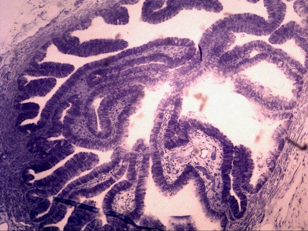
Uterine tube · semithin section, toluidine blue
Labelled structures
This image has no labels – use the caption instead.
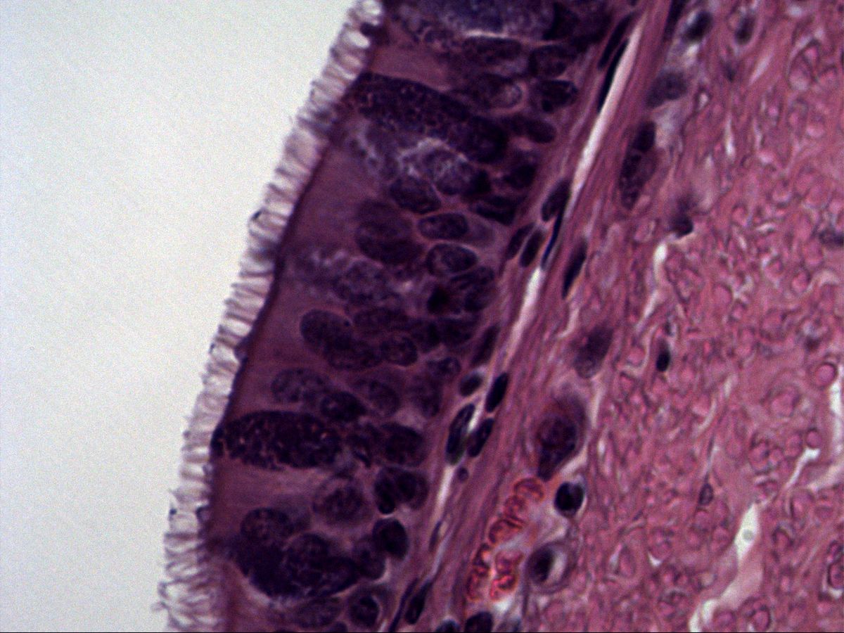
The mucosa of the uterine tube is covered by a simple columnar epithelium made of two cell types: ciliated cells with kinocilia and non-ciliated secretory cells (peg cells). Especially in the ampulla the mucosa forms a labyrinth of tall, branching folds.
In a semithin section (about 0.5–1 µm, resin-embedded, toluidine blue) cell borders, cilia and secretory granules are much sharper than in a paraffin section. This slide is therefore ideal for looking at cilia directly.
How to recognise it
- Simple columnar epithelium
- Ciliated cells with a dense covering of cilia
- Dark secretory cells with bulging apex
- No goblet cells
- Heavily branched mucosal folds
Structures to find
- Cilia
- Ciliated cell
- Secretory (peg) cell
- Mucosal fold
- Lamina propria
- Lumen
- Muscular layer
Structure
The wall of the uterine tube consists of mucosa, muscular layer (inner circular and outer longitudinal smooth muscle) and serosa. The mucosa forms longitudinal, branching tubal folds (plicae tubariae) with a delicate core of cellular lamina propria. The epithelium is simple: ciliated cells with pale cytoplasm, a round-to-oval nucleus and a dense covering of cilia alternate with narrower, darker secretory cells, whose apex bulges dome-like into the lumen and contains secretory granules.
Function
The cilia beat towards the uterus and, together with the peristalsis of the muscular layer, support transport of the oocyte or early embryo. The secretory cells produce tubal fluid that provides nutrients and contributes to sperm capacitation. The epithelium changes during the cycle: under oestrogen cell height and the number of cilia increase, while secretory cells dominate in the luteal phase. After menopause the epithelium becomes lower and loses part of its cilia.
Microscopy tips
At low magnification you recognise the uterine tube by the heavily branched mucosal folds that almost fill the lumen. At high magnification find a stretch of epithelium cut perpendicularly: cilia appear as a fine, pale-blue fringe, secretory cells as dark cells bulging apically without cilia. Important for differential diagnosis: the tubal epithelium is simple and has no goblet cells – unlike the pseudostratified ciliated epithelium of the trachea. In a semithin section the pink hues of H&E are missing; all structures appear in shades of blue.
Clinical relevance
Inflammation of the tube (salpingitis, often caused by chlamydia) can destroy cilia and make the folds adhere. The result is impaired oocyte transport, increasing the risk of ectopic (tubal) pregnancy and infertility. According to current knowledge, many high-grade serous carcinomas of the 'ovary' arise from the epithelium of the fimbriae. For this reason both tubes are now often removed prophylactically during pelvic surgery.
Slide images
Test your knowledge
Assign the correct number in the image to each term.
Tap the requested structure in the image.
Multiple-choice questions on structure and function.
Frequently asked questions
What is a semithin section?
A section about 0.5–1 µm thick from resin-embedded tissue, usually stained with toluidine blue. It shows cell details much more sharply than a standard paraffin section.
Is the tubal epithelium pseudostratified like the trachea?
No, it is simple. Because ciliated and secretory cells differ in width, the nuclei are slightly staggered, but not arranged in clearly separate rows.
What moves the oocyte along the uterine tube?
By ciliary beating towards the uterus and by peristaltic contractions of the smooth muscle, supported by the flow of tubal fluid.
Related topics
Updated: