Simple columnar epithelium
Gallbladder · trichrome
Labelled structures
This image has no labels – use the caption instead.
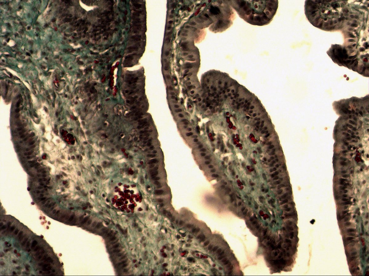
The section of the gallbladder shows a simple columnar epithelium with microvilli for absorption.
The gallbladder mucosa forms numerous branching folds covered by a uniform simple columnar epithelium. The cells are clearly taller than wide, and their elongated nuclei lie in one row in the basal third of the cell. In trichrome staining the connective tissue of the lamina propria stands out green against the grey-violet epithelium.
How to recognise it
- One row of elongated, basally located nuclei
- Cells clearly taller than wide
- Fine apical fringe of microvilli
- No goblet cells
- Branching mucosal folds with loose lamina propria
Structures to find
- Microvilli (brush border)
- Cell borders
- Nuclei
- Basement membrane
- Lamina propria with capillaries
Structure
Each epithelial cell rests on the basement membrane and reaches the lumen. The nucleus is oval to elongated and aligned with the cell axis; because all nuclei lie at about the same level, a single row of nuclei results. Apically the cells carry short microvilli (bundles of actin and fimbrin, about 1–2 µm) that appear as a fine fringe in the light microscope. Laterally the cells are joined by junctional complexes (tight junctions). Beneath the epithelium lies a cell- and vessel-rich lamina propria; the gallbladder has no muscularis mucosae and no submucosa.
Function
The gallbladder stores and concentrates hepatic bile. The epithelium actively absorbs sodium and chloride; water follows osmotically and passes through the intercellular spaces into the capillaries of the lamina propria. The microvilli enlarge the absorptive surface, and tight junctions prevent bile from leaking between the cells into the tissue. In this way bile can be concentrated many times over. In addition, the epithelial cells produce some mucus that protects the surface from aggressive bile acids, and they secrete bicarbonate.
Microscopy tips
At low magnification you see the branching mucosal folds, which can look like villi in section – but goblet cells are absent, which distinguishes the gallbladder from the intestine. At high magnification check: one row of nuclei, cells taller than wide, an apical fringe. Obliquely cut folds are misleading because nuclei seem to lie at several levels, so look for an area where the epithelium is cut perpendicularly. Beneath the mucosa follows a loose wall of smooth muscle bundles and connective tissue.
Clinical relevance
If bile becomes overly concentrated or its composition is disturbed, cholesterol or bilirubin salts precipitate and gallstones form (cholelithiasis). They can cause inflammation of the gallbladder wall (cholecystitis). In chronic inflammation the mucosa pouches deeply into the muscle layer (Rokitansky–Aschoff sinuses). These outpouchings are a typical finding in specimens after cholecystectomy. Long-standing gallstone disease also increases the risk of gallbladder carcinoma.
Slide images
Test your knowledge
Assign the correct number in the image to each term.
Tap the requested structure in the image.
Multiple-choice questions on structure and function.
Frequently asked questions
Why does the gallbladder have no goblet cells?
The epithelial cells themselves produce small amounts of mucus that protect the surface from bile. Their main task, however, is absorbing water and salts, not producing mucus as in the intestine.
Are the gallbladder microvilli a true brush border?
They are shorter and less dense than the brush border of the small intestine or proximal tubule. In the light microscope they appear as a narrow fringe that is often also called a brush border in class.
Which stain does this slide show?
A trichrome stain: collagen fibres appear green or blue, cytoplasm and muscle reddish-grey and erythrocytes bright red.
Related topics
Updated: