Reticular connective tissue
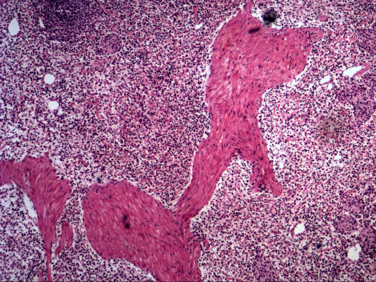
Spleen, perfused · H&E
Labelled structures
This image has no labels – use the caption instead.
Reticular connective tissue forms the three-dimensional framework of lymphoid organs and bone marrow. It consists of stellate, branched reticular cells and the fine reticular fibres (type III collagen) they produce. In the perfused spleen many blood cells have been washed out, so the meshwork stands out more clearly.
How to recognise it
- Loose meshwork with many free cells (mainly lymphocytes)
- Reticular cells: large, pale, oval nuclei and branched processes
- Reticular fibres barely visible in H&E, black with silver stains
- In the spleen: eosinophilic trabeculae of dense connective tissue
- Perfused spleen: empty spaces (sinuses) between the cell cords
Structures to find
- Reticular cells with pale nucleus
- Meshwork (reticular fibres, only faintly visible)
- Free cells within the meshes (lymphocytes)
- Splenic trabeculae
- Washed-out sinuses of the red pulp
Structure
Reticular cells (fibroblastic reticular cells) have a large, pale, oval nucleus and long cytoplasmic processes by which they contact one another. Together with the reticular fibres they ensheathe, they form a loose, sponge-like network. Its meshes contain free cells: lymphocytes, macrophages, plasma cells and, in the red pulp, blood cells.
In the spleen, trabeculae of dense connective tissue also traverse the organ. They arise from the capsule and carry the trabecular vessels.
Function and occurrence
Reticular connective tissue is a supporting scaffold for freely mobile cells and at the same time a site of cell–cell communication: reticular cells produce chemokines, guide lymphocytes and expose them to antigen in the right environment. It occurs in the spleen, lymph nodes, tonsils, Peyer's patches, bone marrow and as the framework of the liver (reticular fibres around the sinusoids). The thymus is an exception: its framework is formed by epithelial reticular cells.
Microscopy tips
In H&E sections reticular fibres are practically invisible – they only show up with silver impregnation (black, 'argyrophilic') or PAS. Focus on the cells instead: among the many small, round, dark lymphocyte nuclei you will find occasional larger, paler oval nuclei with faint, process-rich cytoplasm – these are reticular cells. At low power the strongly eosinophilic trabeculae help with orientation. Washed-out empty spaces correspond to splenic sinuses or the red pulp meshwork.
Clinical relevance
In bone marrow fibrosis (e.g. myelofibrosis) reticular fibres increase markedly; the degree of fibrosis is graded on silver stains. Clinically this often presents as a 'dry tap', in which no marrow can be aspirated. In lymph nodes and spleen the reticular framework is often destroyed or displaced by lymphoma, which is used diagnostically in reticulin stains. In the liver, too, collapse of the reticulin network indicates loss of hepatocytes.
Slide images
Test your knowledge
Assign the correct number in the image to each term.
Tap the requested structure in the image.
Multiple-choice questions on structure and function.
Frequently asked questions
Why can't reticular fibres be seen in H&E sections?
They are very thin and coated with glycoproteins, so they hardly take up eosin. With silver impregnation they appear black, hence the term 'argyrophilic fibres'.
Why is the spleen perfused for this preparation?
Perfusion removes the numerous red blood cells of the red pulp. This makes the meshwork of reticular cells and the sinuses more clearly visible.
Is the framework of the thymus also reticular connective tissue?
No. The thymus has a framework of epithelial reticular cells joined by desmosomes, which do not form reticular fibres.
Related topics
Updated: