Tubuloacinar gland
Submandibular gland · H&E
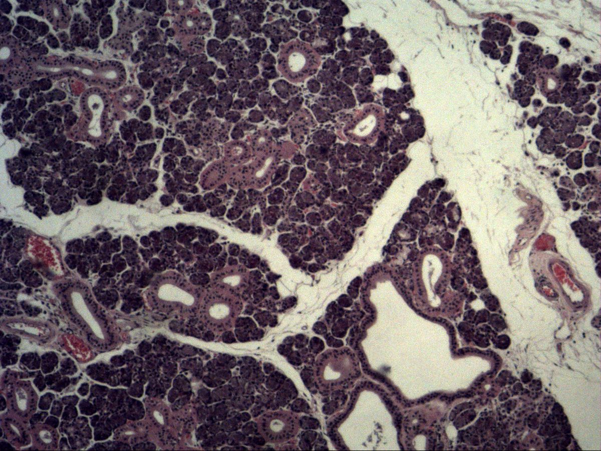
Labelled structures
This image has no labels – use the caption instead.
This is a mixed gland: a seromucous gland. This means that you find more serous than mucous glandular units, which can be seen here from the end pieces. A demilune and an excretory duct are also visible. The ducts are divided into intercalated ducts and striated ducts (larger lumen).
The submandibular gland is a compound (branched) tubuloacinar gland: serous acini and mucous tubules drain into a branched duct system of intercalated ducts, striated ducts and interlobular excretory ducts. Connective tissue septa divide the organ into lobules. Its well-developed, eosinophilic striated ducts make it a textbook specimen.
How to recognise it
- Mixed gland: mainly serous acini with pale mucous tubules in between
- Serous demilunes on mucous end pieces
- Numerous eosinophilic striated ducts with a round lumen
- Lobulation by connective tissue septa
- Interlobular excretory ducts within the septa
Structures to find
- Serous end piece
- Mucous end piece (flat nucleus)
- Serous demilune
- Intercalated duct
- Striated duct (intralobular duct)
- Interlobular excretory duct
- Basement membrane
Structure
Serous end pieces (acini) are round with a very narrow lumen; their pyramidal cells have a round, basal nucleus and basophilic cytoplasm with apical granules. Mucous end pieces are tubular with a wide lumen; their cells look pale and foamy, with the nucleus flattened against the base. Serous cells sitting like a cap on a mucous tubule form the serous demilune (demilune of von Ebner/Giannuzzi). Myoepithelial cells embrace the end pieces. The duct system begins with short intercalated ducts (flat to cuboidal epithelium), followed by long striated ducts with tall columnar, strongly eosinophilic epithelium and basal striations, and drains into interlobular ducts within the septa.
Function
The submandibular gland produces most of the saliva at rest. Serous cells form a watery, enzyme-rich secretion (including α-amylase and lysozyme), mucous cells a viscous, mucin-containing mucus. The end pieces produce a primary saliva isotonic with plasma. The striated ducts reabsorb Na⁺ and Cl⁻ and secrete K⁺ and HCO₃⁻; the basal striations correspond to infoldings of the plasma membrane with numerous mitochondria that power this active transport. As a result, the saliva becomes hypotonic. Myoepithelial cells squeeze the secretion out of the end pieces.
Microscopy tips
In the overview you can see lobules separated by pale connective tissue septa. Then look for the mixture: small, dark serous acini predominate, with pale mucous tubules in between. The striated ducts stand out within the lobules as pink, ring-shaped cross-sections with a round lumen – they are particularly numerous in the submandibular gland. At high magnification look for the crescent-shaped cap of dark serous cells on mucous tubules. Differential diagnosis: the parotid is purely serous, the sublingual gland mainly mucous with few striated ducts; the pancreas has no striated ducts but has islets of Langerhans.
Clinical relevance
The long, ascending excretory duct (submandibular duct) and the more viscous, calcium-rich saliva explain why salivary stones (sialolithiasis) form most often in the submandibular gland. A painful swelling during meals is typical. In Sjögren's syndrome, lymphocytic inflammation destroys the end pieces and causes a dry mouth. Salivary gland tumours such as pleomorphic adenoma can also occur here. When the gland is removed surgically, the closely related marginal mandibular branch of the facial nerve is at risk.
Slide images
Test your knowledge
Assign the correct number in the image to each term.
Tap the requested structure in the image.
Multiple-choice questions on structure and function.
Frequently asked questions
Why are serous end pieces darker than mucous ones?
Serous cells contain plenty of rough ER and protein granules that take up haematoxylin and eosin. Mucous cells are filled with poorly staining mucins and look pale.
What are the basal striations of striated ducts?
They are deep infoldings of the basal plasma membrane with mitochondria lined up between them. They supply the energy for the ion transport that makes saliva hypotonic.
How do I tell the submandibular gland from the parotid?
The parotid is purely serous. If you find pale mucous tubules and serous demilunes, the section is from the submandibular gland.
Related topics
Updated: