Spermatogenesis
Testis · trichrome
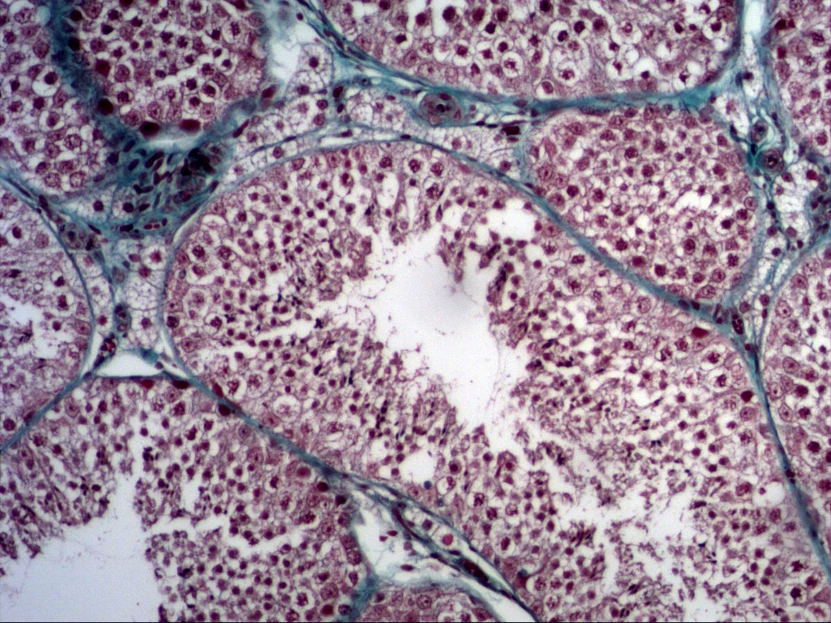
Labelled structures
This image has no labels – use the caption instead.
The seminiferous tubules, in which the sperm are produced, are clearly visible. Development proceeds from the base towards the lumen: spermatogonia, spermatocytes and spermatids. The Sertoli cells form the epithelium and at the same time the blood–testis barrier (tight junctions). The Leydig cells lie in the connective tissue and are responsible for testosterone production.
The germinal epithelium of the convoluted seminiferous tubules is a special epithelium: it consists of stationary Sertoli cells and the germ cells, which move between them from the basement membrane towards the lumen while passing through mitosis, meiosis and transformation into spermatozoa (spermiogenesis). In a tubule cross-section you therefore see all stages side by side.
How to recognise it
- Round tubules with a multilayered germinal epithelium
- Maturation gradient from basal (spermatogonia) to luminal (spermatids)
- Large primary spermatocytes with thread-like chromatin
- Sperm heads and flagella in the lumen
- Groups of Leydig cells in the interstitium
Structures to find
- Spermatogonia
- Primary spermatocyte
- Spermatids
- Spermatozoa
- Sertoli cells (prominent nucleoli)
- Lamina propria
- Leydig cells
Structure
Each tubule is surrounded by a lamina propria made of basement membrane, collagen fibres and contractile myofibroblasts. Directly on the basement membrane lie the spermatogonia: small, round cells with a dark nucleus. Above them are the primary spermatocytes, the largest germ cells, with a large nucleus and thread-like chromatin (long prophase of meiosis I). Secondary spermatocytes exist only briefly and are rarely seen. Towards the lumen lie the small, round early spermatids and the elongated late spermatids with dark, elongated heads. The Sertoli cells extend from the basement membrane to the lumen; their pale, often triangular nucleus with a prominent nucleolus lies basally. In the interstitium the Leydig cells lie in groups around capillaries.
Function
Spermatogonia multiply by mitosis and maintain the stem cell pool. They give rise to primary spermatocytes, which become secondary spermatocytes in meiosis I and haploid spermatids in meiosis II. During spermiogenesis the spermatids form the acrosome and flagellum, condense their chromatin and shed most of their cytoplasm. In humans the whole process takes about 64–74 days. Sertoli cells nourish the germ cells, form the blood–testis barrier and secrete androgen-binding protein and inhibin. Leydig cells produce testosterone under the influence of LH.
Microscopy tips
At low magnification you see densely packed, round tubule cross-sections with a multilayered epithelium and narrow interstitium between them (green in trichrome). Then work from the outside inwards: basement membrane – spermatogonia – large spermatocytes with chromatin threads – small round spermatids – sperm heads and flagella in the lumen. Sertoli cells are best identified by their pale nucleus with a large nucleolus near the base. Leydig cells are large, eosinophilic cells with a round nucleus in the interstitium. Not every tubule shows all stages – look at several.
Clinical relevance
In cryptorchidism (undescended testis) the higher body temperature impairs spermatogenesis; if untreated, infertility and an increased risk of germ cell tumours result. These testicular tumours, especially seminomas, are the most common solid tumour in young men. In Klinefelter syndrome (47,XXY) the tubules are hyalinised and largely devoid of germ cells. The blood–testis barrier protects germ cells, which only form after puberty, from the body's own immune system; after injury, autoantibodies against sperm may therefore develop.
Slide images
Test your knowledge
Assign the correct number in the image to each term.
Tap the requested structure in the image.
Multiple-choice questions on structure and function.
Frequently asked questions
Why are secondary spermatocytes hardly ever seen?
They enter meiosis II immediately after meiosis I and exist for only a few hours. Primary spermatocytes, in contrast, remain in prophase I for about three weeks.
How do I recognise a Sertoli cell?
By its pale, irregular to triangular nucleus with a large, dark nucleolus near the basement membrane. The cell boundaries themselves are barely visible under the light microscope.
Where exactly is the blood–testis barrier located?
It is formed by tight junctions between neighbouring Sertoli cells and separates a basal compartment (spermatogonia) from an adluminal compartment (spermatocytes, spermatids).
Related topics
Updated: