Oocytes and follicle maturation
Ovary · H&E
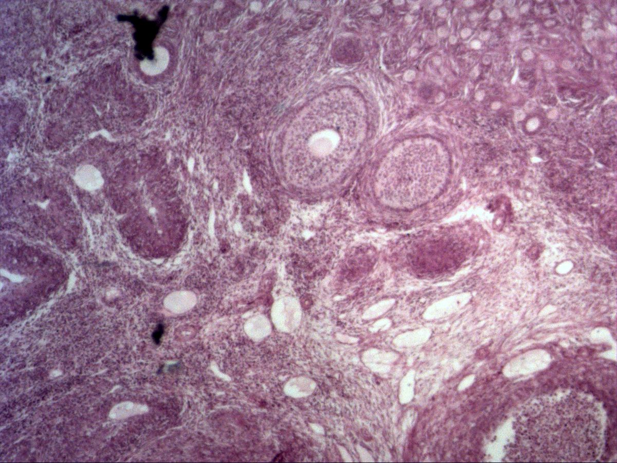
Labelled structures
This image has no labels – use the caption instead.
The ovary contains the ovarian follicles ('little vesicles'). Their development is divided into the following stages: primordial follicle (single layer of flat epithelium), primary follicle (single layer of cuboidal epithelium, zona pellucida).
These are followed by the secondary follicle, with a multilayered follicular epithelium (stratum granulosum) and a developing theca from the surrounding stroma, and the tertiary follicle, in which a fluid-filled cavity, the antrum, forms between the granulosa cells. The oocyte then lies eccentrically on a mound of granulosa cells (cumulus oophorus). The mature tertiary follicle ready to ovulate is called the Graafian follicle. In all stages the oocyte is arrested in prophase of the first meiotic division (dictyotene).
How to recognise it
- Cortex with follicles in a cell-rich, spindle-cell stroma
- Large oocytes with a pale nucleus and nucleolus
- Follicular epithelium flat, cuboidal or multilayered depending on stage
- Eosinophilic zona pellucida around the oocyte
- Tertiary follicle with a fluid-filled antrum
Structures to find
- Primordial follicle
- Primary follicle
- Secondary follicle
- Tertiary follicle with antrum
- Oocyte with nucleus and nucleolus
- Zona pellucida
- Follicular cells and basement membrane
Structure
The ovary is divided into a cortex, with follicles in a cell-rich, spindle-cell stroma, and a vascular medulla. On the outside lies a simple flat to cuboidal surface epithelium, beneath it the tough tunica albuginea. Primordial follicles lie in large numbers just below the surface: an oocyte with a large, pale nucleus surrounded by a few flat follicular cells. In the primary follicle the cells are cuboidal, and the eosinophilic zona pellucida appears between oocyte and epithelium. From the secondary follicle onwards, the stroma around the follicle differentiates into a cell-rich, hormone-producing theca interna and a more fibrous theca externa; both are separated from the granulosa by a basement membrane.
Function
At birth the ovary contains about 1–2 million primordial follicles, at puberty around 400,000; only about 400 of these ovulate during a lifetime. In each cycle a cohort of follicles grows, usually only one dominant follicle reaches ovulation, and the rest become atretic. Theca interna cells produce androgens under the influence of LH, which granulosa cells aromatise into oestrogens under the influence of FSH. Shortly before ovulation the oocyte completes meiosis I and arrests again in metaphase II. After ovulation the remnant of the follicle becomes the corpus luteum.
Microscopy tips
First find the surface of the ovary and work inwards: beneath the surface epithelium lie nests of small primordial follicles, with the larger stages deeper. Classification depends on the shape and number of follicular cell layers and on the presence of an antrum. The oocyte is not hit in every section – a follicle without an oocyte in the image is not a mistake but a tangential cut. You can recognise atretic follicles by a shrunken oocyte, a folded zona pellucida and disintegrating granulosa cells. The stroma differs from smooth muscle by its whorled arrangement of spindle-shaped cells.
Clinical relevance
In polycystic ovary syndrome (PCOS) many follicles start to mature without ovulating, resulting in raised androgens, cycle disturbances and infertility. The number of remaining follicles – the ovarian reserve – can be estimated from anti-Müllerian hormone produced by granulosa cells. Chemotherapy and radiotherapy can destroy the follicle pool, so ovarian tissue is cryopreserved beforehand in young patients. At menopause the pool is exhausted.
Slide images
Test your knowledge
Assign the correct number in the image to each term.
Tap the requested structure in the image.
Multiple-choice questions on structure and function.
Frequently asked questions
How do I tell a primary from a secondary follicle?
The primary follicle has a single layer of cuboidal follicular cells. In the secondary follicle the epithelium is multilayered and a theca begins to form.
At which stage of meiosis is the oocyte in the follicle?
It has been arrested in prophase I (dictyotene) since fetal life. Meiosis I is only completed shortly before ovulation.
Why do some follicles appear without an oocyte?
The oocyte is small relative to the follicle and is only hit if the section passes through its centre. Peripheral cuts show only granulosa, antrum and theca.
Related topics
Updated: