Gastrulation – early germ layers
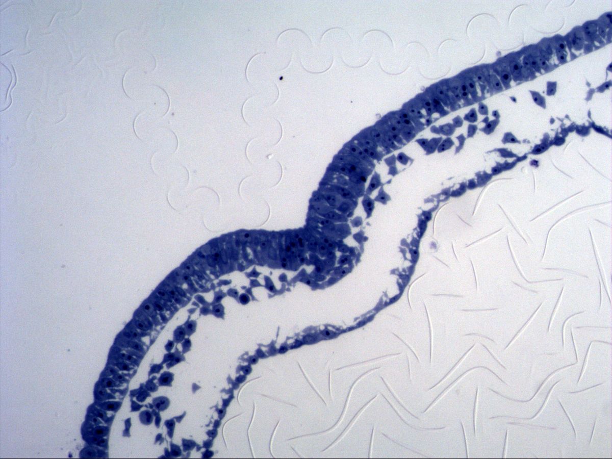
Primitive streak · semithin section
Labelled structures
This image has no labels – use the caption instead.
Gastrulation is the step in which the bilaminar embryonic disc (epiblast and hypoblast) gives rise to the three germ layers: ectoderm, mesoderm and endoderm. In the midline of the epiblast the primitive streak forms, with a furrow, the primitive groove. There, epiblast cells detach from the epithelium, migrate downwards and spread out as loose mesenchyme between the layers.
The specimen comes from a pig embryo and is a semithin section (plastic embedding, about 1 µm thick) stained with Richardson's stain (methylene blue/azure II). Cell boundaries, nuclei and nucleoli are therefore particularly sharp – ideal for following the transformation of epithelial cells into mesenchymal cells.
How to recognise it
- Thick, columnar epiblast layer
- Thin hypoblast layer of flat cells
- Loose, star-shaped mesenchymal cells between the layers
- Thickened epiblast with emigrating cells at the primitive streak
- Semithin section: sharp cell boundaries, blue stain, distinct nucleoli
Structures to find
- Epiblast
- Hypoblast
- Mesenchyme
- Primitive streak / primitive groove
- Bottle cells (EMT)
Structure
The epiblast is a tall columnar, pseudostratified epithelium with crowded nuclei and forms the thick upper layer. Beneath it lies the hypoblast, a thin layer of flat cells facing the yolk sac. In the region of the primitive streak the epiblast is thickened; here there are wedge-shaped bottle cells with a narrow apical neck and a broadened basal cell body. Between epiblast and hypoblast lie star-shaped, loosely scattered mesenchymal cells with processes and much intercellular space. The basement membrane beneath the epiblast is interrupted at the primitive streak.
Function
During epithelial–mesenchymal transition (EMT), epiblast cells lose their apicobasal polarity and their cell–cell contacts (down-regulation of E-cadherin), constrict apically (bottle cells), break through the basement membrane and migrate away as mesenchyme. The first cells to ingress replace the hypoblast and form the definitive endoderm; the following cells form the intraembryonic mesoderm between the layers. The remaining epiblast becomes the ectoderm. Cells migrating through the primitive node form the notochord. All three germ layers are thus derived from the epiblast.
Microscopy tips
At low magnification orient yourself by the thick, dark blue epiblast. The thin, often discontinuous row of cells opposite is the hypoblast, with individual mesenchymal cells in between. Then look for the place where the epiblast is thickened, loses its smooth lower edge and releases cells downwards – this is the primitive streak. At high magnification look for wedge-shaped bottle cells and for cells that are just detaching from the epithelium. In the semithin section nuclei show distinct, deep blue nucleoli; mitoses are common in the epiblast.
Clinical relevance
Disturbances of gastrulation have far-reaching consequences. If remnants of the primitive streak persist, sacrococcygeal teratomas containing tissue from all three germ layers can develop. Insufficient mesoderm formation in the caudal region leads to caudal dysgenesis (sirenomelia). EMT is also a key mechanism of tumour metastasis: carcinoma cells reactivate this embryonic programme, lose E-cadherin and become invasive. Teratogens such as alcohol also act during this early, highly sensitive phase.
Slide images
Test your knowledge
Assign the correct number in the image to each term.
Tap the requested structure in the image.
Multiple-choice questions on structure and function.
Frequently asked questions
What is the difference between epiblast and ectoderm?
The epiblast is the upper layer of the bilaminar disc and the source of all three germ layers. Only the epiblast remaining after gastrulation is called ectoderm.
What are bottle cells?
Epiblast cells that constrict strongly at their apex and thus become wedge- or bottle-shaped. They initiate the ingression of cells at the primitive streak.
Why is a semithin section used?
The roughly 1 µm thin plastic section shows cell boundaries and shapes much more sharply than a paraffin section. This makes it easy to follow individual migrating cells.
Related topics
Updated: