Lymphoid follicles and lymphoid tissue
Lymphoid organs · H&E
Labelled structures
This image has no labels – use the caption instead.
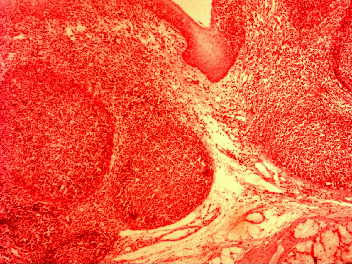
Lymphoid tissue consists of a framework of reticular cells and fibres whose meshes are densely packed with lymphocytes. It occurs either diffusely or condensed into round nodules. These lymphoid follicles (lymphoid nodules) are B-cell zones: a primary follicle is uniformly dark, whereas a secondary follicle, after antigen contact, has a pale germinal centre and a dark mantle zone.
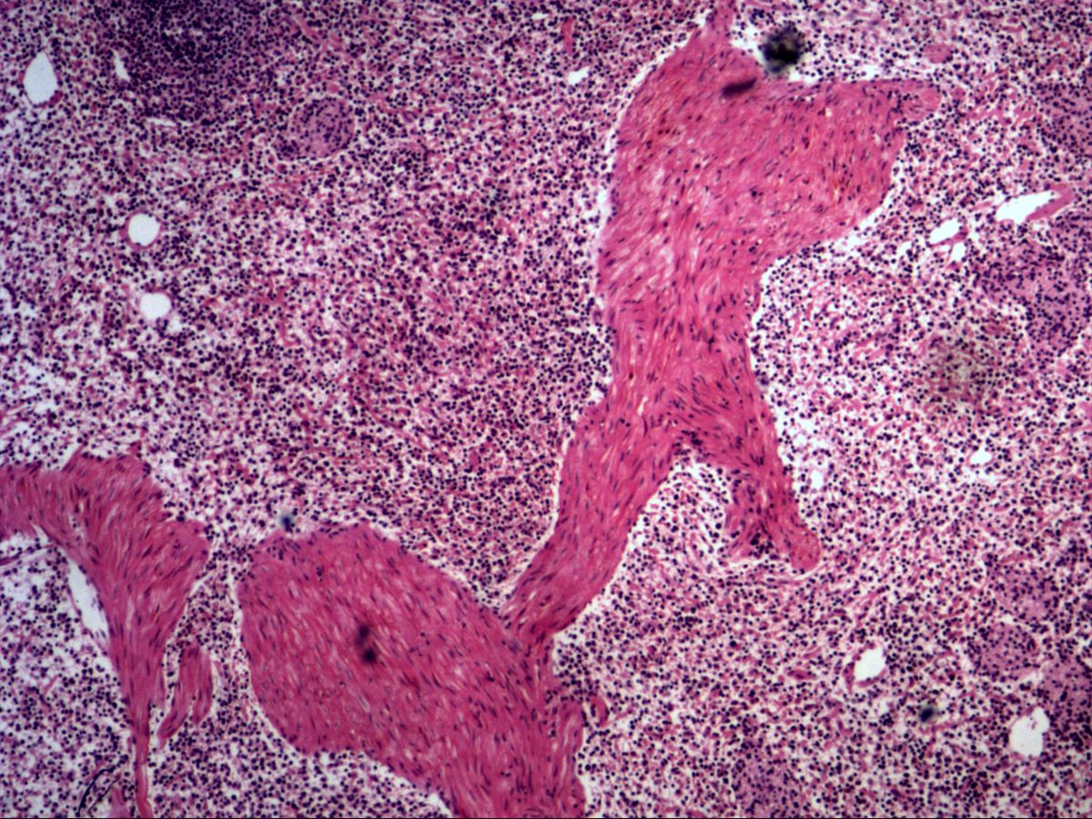
The older H&E images shown here come from different lymphoid organs and have not been reliably assigned. They probably show lymph node and tonsil, lymphocytes at high magnification and, for comparison, thymus with Hassall’s corpuscles. Read the captions as reasoned assessments.
How to recognise it
- Densely packed, small, round, dark nuclei (lymphocytes)
- Round, dark nodules (follicles) within diffuse lymphoid tissue
- Secondary follicle: pale germinal centre with a dark mantle zone
- Fine reticular framework with vessels between the lymphocytes
- Organ-specific features: capsule and sinus, crypt epithelium or central artery
- Thymus: lobules with Hassall’s corpuscles but no follicles
Structures to find
- Primary follicle
- Secondary follicle
- Germinal centre
- Mantle zone
- Diffuse lymphoid tissue
- Capsule (lymph node)
- Small lymphocytes
- Hassall’s corpuscles (thymus, for comparison)
Structure
- Primary follicle: a round, uniformly dark cluster of small, resting (naive) B lymphocytes on a network of follicular dendritic cells.
- Secondary follicle: develops after antigen contact. Its centre is the pale germinal centre with large proliferating centroblasts (dark zone) and centrocytes (light zone), follicular dendritic cells and macrophages containing phagocytosed debris (tingible body macrophages).
- Mantle zone: a dense, dark rim of small, non-activated B cells that caps the germinal centre.
- Marginal zone: a paler rim outside the mantle zone, especially prominent in the spleen.
Between the follicles lies diffuse lymphoid tissue containing mainly T cells and high endothelial venules.
Occurrence and function
Lymphoid follicles occur in all secondary lymphoid organs: in the cortex of lymph nodes, in the white pulp of the spleen, in the tonsils beneath the crypt epithelium and in mucosa-associated lymphoid tissue (MALT), for example Peyer’s patches of the ileum and the appendix. In the germinal centre, antigen-activated B cells proliferate, modify their antibodies by somatic hypermutation and are selected for high affinity (affinity maturation). This gives rise to plasma cells and memory B cells. The thymus, by contrast, is a primary lymphoid organ for T-cell maturation and normally has no lymphoid follicles.
Microscopy tips
At low power lymphoid tissue is recognised by densely packed, dark blue nuclei without obvious tissue architecture. Then look for structures that identify the organ:
- Lymph node: connective tissue capsule, subcapsular sinus beneath it, follicles in the cortex, medullary cords and sinuses
- Tonsil: surface epithelium with deep crypts, follicles directly beneath
- Spleen: white pulp with central artery embedded in red pulp, trabeculae with vessels
- Thymus: lobules with dark cortex and pale medulla, Hassall’s corpuscles, no follicles
At high power small lymphocytes are round with dense chromatin and a thin rim of cytoplasm.
Clinical relevance
During infections, lymph nodes and tonsils swell because numerous secondary follicles with large germinal centres develop (reactive follicular hyperplasia). Such a lymph node is usually tender and freely mobile. Follicular lymphoma arises from germinal centre cells; densely packed, uniform follicles without tingible body macrophages efface the normal architecture. Painless, firm and matted lymph nodes therefore always require investigation. Recurrent tonsillitis may make tonsillectomy necessary.
Slide images
Test your knowledge
Assign the correct number in the image to each term.
Tap the requested structure in the image.
Multiple-choice questions on structure and function.
Frequently asked questions
What is the difference between a primary and a secondary follicle?
A primary follicle is a uniformly dark cluster of resting B cells. A secondary follicle develops after antigen contact and has a pale germinal centre surrounded by a dark mantle zone.
Why is the germinal centre paler than the mantle zone?
The germinal centre contains large activated B cells with paler, open chromatin and more cytoplasm, plus follicular dendritic cells and macrophages. The mantle zone consists of densely packed small lymphocytes with dark nuclei.
Does the thymus have lymphoid follicles?
Normally not. The thymus is a primary lymphoid organ divided into lobules with dark cortex and pale medulla and is identified by Hassall’s corpuscles in the medulla. Follicles in the thymus occur, for example, in myasthenia gravis.
Related topics
Updated: